Comparative Study
. 2007 Apr 17;104(16):6590-5.
doi: 10.1073/pnas.0701776104. Epub 2007 Apr 9.

Direct and adaptor-mediated substrate recognition by an essential AAA+ protease

Affiliations
Comparative Study

Direct and adaptor-mediated substrate recognition by an essential AAA+ protease

Peter Chien et al. Proc Natl Acad Sci U S A. .

Abstract

Regulated proteolysis is required to execute many cellular programs. In Caulobacter crescentus, timely degradation of the master regulator CtrA by ClpXP protease is essential for cell-cycle progression and requires the colocalization of CtrA and RcdA. Here, we establish a biochemical framework to understand regulated proteolysis in C. crescentus and show that RcdA is not an adaptor for CtrA degradation. CtrA is rapidly degraded without RcdA and is recognized with an affinity comparable with the best ClpXP substrates. In contrast, SspBalpha, the alpha-proteobacterial homolog of SspB, functions as an adaptor to enhance degradation of specific substrates. Cargo-free SspBalpha is also itself a substrate of ClpXP-mediated proteolysis. Thus, our analysis (i) reveals the consequences of both direct and adaptor-stimulated recognition in mediating substrate specificity in vitro, (ii) reveals a potential regulatory role of controlled adaptor stability, and (iii) suggests that cell-cycle regulation of CtrA stability depends on repression of its intrinsic degradation rather than adaptor-mediated enhancement.


Conflict of interest statement

The authors declare no conflict of interest.

Figures

Fig. 1.
Fig. 1.
Caulobacter ClpXP is an active protease. (A Upper) Diagram depicting stages of the cell cycle when CtrA is degraded and when CtrA/RcdA are colocalized. (A Lower) Sequences of E. coli ssrA peptide tag, C. crescentus ssrA peptide tag, and C-terminal residues of CtrA. Regions critical for ClpX recognition are highlighted. (B) GFP with C. crescentus ssrA peptide fused to the C-terminal end is a substrate for degradation by ClpXP (CCssrA). Mutation of C-terminal alanine residues to aspartate residues (filled squares) eliminates degradation. Degradation depends on ATP (filled triangles), and C. crescentus ClpX does not function with E. coli ClpP (open circles). At the indicated times, ATP regenerating mix or C. crescentus ClpP was added to reactions lacking these components. Reactions consist of 0.3 μM ClpX, 0.8 μM ClpP, and 2 μM substrate. (C) SDS/PAGE gel of reactions showing that CtrA is degraded in vitro (CtrA) and requires ATP (CtrA no ATP). Mutating C-terminal alanine residues to aspartate residues eliminates degradation (CtrA-DD). The band visible between ClpX and CtrA is creatine kinase (labeled CK), a component of the ATP regeneration system. Reactions consist of 0.3 μM ClpX, 0.8 μM ClpP, and 1 μM CtrA plus an ATP regeneration system when indicated. (D) Initial rates of CtrA degradation by ClpXP as a function of substrate concentration. The fit is to the Michaelis–Menten equation with a KM of 1.2 μM and Vmax of 3.9 molecules degraded per ClpX6 per min. (E) Truncated CtrA lacking the N-terminal 116 residues is degraded in vitro by ClpXP wild type (Left) but not when the C-terminal AA motif is mutated to DD (Right). Reactions consist of 0.2 μM ClpX, 0.4 μM ClpP, and 8 μM truncated CtrA plus the ATP regeneration mix.
Fig. 2.
Fig. 2.
RcdA does not affect CtrA degradation. (A) Low (equimolar) or high (5-fold excess) amounts of RcdA do not affect CtrA degradation by ClpXP. Fraction of remaining CtrA (y axis) is plotted versus time; concentrations in the reactions were 0.5 μM CtrA, 0.5 μM RcdA, 0.3 μM ClpX, and 0.8 μM ClpP (low RcdA) and 1.0 μM CtrA, 5 μM RcdA, 0.2 μM ClpX, and 0.4 μM ClpP (high RcdA). Lines are fits to exponential decays. (B) RcdA does not bind ClpX effectively as assayed by retention during filtration through a 100-kDa cutoff filter. ClpX hexamers are retained on these filters, whereas smaller proteins such as RcdA flow through. Shown are total samples before filtration (T), flow-through after filtration (F), and retentate (R) for either RcdA (0.5 μM) alone or RcdA plus ClpX6 (0.4 μM). Binding was performed in 1× PD buffer (see Materials and Methods) supplemented with 100 mM KCl and 2 mM ATPγS. (C) Purified RcdA is natively folded. Wavelength center of mass (in nanometers) of emission spectrum is plotted as a function of denaturant concentration.
Fig. 3.
Fig. 3.
SspBα is a functional homolog of SspB. (A) SspB organization highlighting the substrate binding domain, the linker region, and the peptide module responsible for ClpX interaction. A homolog of SspB is conserved in the α-proteobacteria family with a domain organization similar to that in characterized SspBs. Alignments were generated by using Jalview (36). (B) SspBα stimulates degradation of GFP-CCssrA by ClpXP. Curves are fits to the Michaelis–Menten equation. (Inset) SspBα binds the C. crescentus ssrA tag, monitored by using fluorescence anisotropy. The curve is a fit to an equilibrium binding equation with a dissociation constant of 0.3 ± 0.02 μM. (C) C-terminal peptide of SspBα is necessary for enhanced degradation of ssrA. Fluorescence of GFP-CCssrA was monitored in reactions containing full-length SspBα (SspBα) or SspBα lacking 5 residues from its C terminus (SspBαΔ5). (D) SspBα is a substrate for ClpXP degradation and is stabilized by substrate binding. Shown are SDS/PAGE of reactions with SspBα, ClpXP, and GFP-CCssrA where indicated.
Fig. 4.
Fig. 4.
Adaptors can positively and negatively regulate degradation. In the absence of an adaptor, both GFP-CCssrA and CtrA are readily degraded. Addition of SspBα enhances GFP-CCssrA degradation but also inhibits degradation of CtrA. (Upper) SDS/PAGE gels of reactions with (Right) and without (Left) SspBα. (Lower) Quantification of gels with fraction remaining of CtrA (Left) or GFP-CCssrA (Right) plotted versus time. Reactions consist of 0.3 μM ClpX, 0.8 μM ClpP, 5 μM CtrA, and 5 μM GFP-CCssrA with addition of 1 μM SspBα where indicated.
Fig. 5.
Fig. 5.
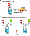
Mechanistic models for regulated degradation of CtrA. (A) Degradation of CtrA may be regulated by relief of an inhibitor that occludes the C-terminal degradation motif of CtrA. Loss of inhibition would be caused by RcdA binding and is dependent on the N-terminal domain of CtrA. (B) Regulation of CtrA degradation could also be controlled, at least in part, by an inhibitor that blocks ClpXP activity. In this case, GFP-CCssrA and CtrA are used as examples of adaptor-dependent and -independent substrates.

Similar articles

Cited by

References

    1. Baker TA, Sauer RT. Trends Biochem Sci. 2006;31:647–653. - PMC - PubMed
    1. Sauer RT, Bolon DN, Burton BM, Burton RE, Flynn JM, Grant RA, Hersch GL, Joshi SA, Kenniston JA, Levchenko I, et al. Cell. 2004;119:9–18. - PMC - PubMed
    1. Jenal U, Hengge-Aronis R. Curr Opin Microbiol. 2003;6:163–172. - PubMed
    1. Dougan DA, Mogk A, Zeth K, Turgay K, Bukau B. FEBS Lett. 2002;529:6–10. - PubMed
    1. Ortega J, Singh SK, Ishikawa T, Maurizi MR, Steven AC. Mol Cell. 2000;6:1515–1521. - PubMed

Publication types

MeSH terms

LinkOut - more resources