. 2007 Apr 24;104(17):7157-62.
doi: 10.1073/pnas.0606974104. Epub 2007 Apr 9.

Mutation of the MAP kinase DYF-5 affects docking and undocking of kinesin-2 motors and reduces their speed in the cilia of Caenorhabditis elegans

Affiliations

Mutation of the MAP kinase DYF-5 affects docking and undocking of kinesin-2 motors and reduces their speed in the cilia of Caenorhabditis elegans

Jan Burghoorn et al. Proc Natl Acad Sci U S A. .

Abstract

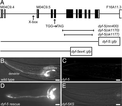
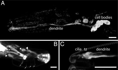
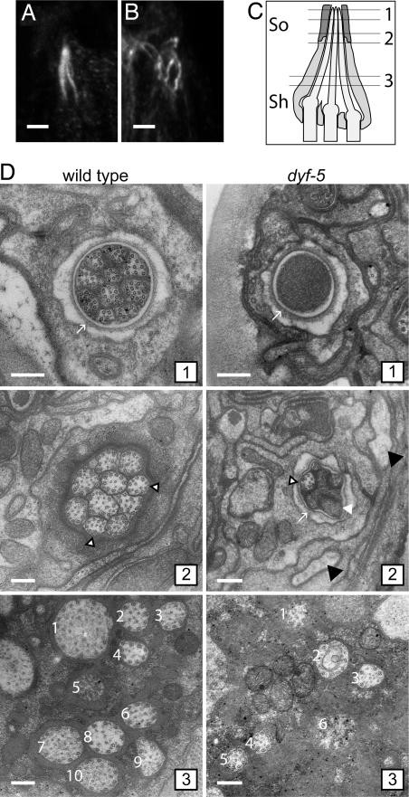
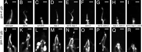
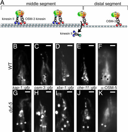
In the cilia of the nematode Caenorhabditis elegans, anterograde intraflagellar transport (IFT) is mediated by two kinesin-2 complexes, kinesin II and OSM-3 kinesin. These complexes function together in the cilia middle segments, whereas OSM-3 alone mediates transport in the distal segments. Not much is known about the mechanisms that compartmentalize the kinesin-2 complexes or how transport by both kinesins is coordinated. Here, we identify DYF-5, a conserved MAP kinase that plays a role in these processes. Fluorescence microscopy and EM revealed that the cilia of dyf-5 loss-of-function (lf) animals are elongated and are not properly aligned into the amphid channel. Some cilia do enter the amphid channel, but the distal ends of these cilia show accumulation of proteins. Consistent with these observations, we found that six IFT proteins accumulate in the cilia of dyf-5(lf) mutants. In addition, using genetic analyses and live imaging to measure the motility of IFT proteins, we show that dyf-5 is required to restrict kinesin II to the cilia middle segments. Finally, we show that, in dyf-5(lf) mutants, OSM-3 moves at a reduced speed and is not attached to IFT particles. We propose that DYF-5 plays a role in the undocking of kinesin II from IFT particles and in the docking of OSM-3 onto IFT particles.


Conflict of interest statement

The authors declare no conflict of interest.

Figures

Fig. 1.
Fig. 1.
dyf-5 encodes a putative protein kinase. (A) Schematic representation of the dyf-5 gene structure (M04C9.5). Coding exons are indicated as black boxes; 5′ and 3′ UTR are depicted as gray boxes. Exons of the flanking genes are indicated as open boxes. The predicted X-box is indicated. Arrows indicate the position of the G-to-A substitution in dyf-5(mn400) animals, and the regions deleted in the ok1170 and ok1177 alleles. The two dyf-5::gfp fusion constructs have been indicated. (B–E) DiI dye-filling of wild-type (B), dyf-5(mn400) (C), dyf-5(ok1170) gjEx824(dyf-5) rescue strain (D), and dyf-5XS(gjEx817) (E) animals. Both dyf-5(lf) and dyf-5(XS) animals are dye-filling-defective. (Scale bars, 20 μm.) Anterior is to the left.
Fig. 2.
Fig. 2.
dyf-5::gfp expression pattern. (A) Expression of dyf-5::gfp in the dendrites and cell bodies of many neurons, including amphid sensory neurons in the head. (B) Strong dyf-5::gfp expression in many neurons in the male tail. Cell bodies are indicated with arrowheads, expression in the sensory rays is indicated with arrows, and autofluorescence of the spicule and the posterior end of the fan is indicated with asterisks. (C) Strong dyf-5::gfp expression in the tip of the head in dendrites and transition zone (tz) and weakly in cilia. [Scale bars, 20 μm (A and B) or 10 μm (C).] Anterior is to the left.
Fig. 3.
Fig. 3.
Mutation of dyf-5 affects cilia length and morphology. (A and B) Anti-tubulin immunostaining of wild-type (A) and dyf-5(ok1177) (B) animals. (Scale bar, 2 μm.) Anterior is up. (C) Schematic representation of three of the amphid channel cilia embedded in the sheath cell (sh) and the socket cell (so). The approximate positions of the EM cross-sections in D have been indicated. (D) EM cross-section of a wild-type (Left 1–3) and dyf-5(lf) (Right 1–3) animal (1). Sections through the socket cell (arrow), showing the distal segments of the 10 channel cilia that contain singlet microtubules in the wild-type animal and the channel filled with electron-dense material in the dyf-5(lf) animal (2). Ten channel cilia embedded in the sheath cell are present in the wild-type animal. Most cilia contain doublet microtubules; some contain singlets (open arrowheads). Section of a dyf-5(lf) animal through the socket cell (arrow), showing some cilia with singlet microtubules (open arrowhead) and some cilia filled with electron-dense material (white arrowhead). Black arrowheads indicate oblique sections through cilia (3). Sections through the middle segments close to the base of the cilia. In the wild-type animal, 10 channel cilia (numbered 1–10) embedded in the sheath cell are present, containing doublet microtubules. In the dyf-5(lf) animal, six channel cilia (–6) embedded in the sheath cell could be identified, containing doublet microtubules. The cilia are more dispersed than in wild-type animals. (Scale bars, 200 nm.)
Fig. 4.
Fig. 4.
Mutation of dyf-5 affects cilia length. Visualization of ASI cilia structures using gpa-4::gfp (A–I) and ADL, ASH, and ASK cilia morphology using gpa-15::gfp (J–R) in wild-type (A and J), dyf-5(ok1177) (B and K), dyf-5XS(gjIs828) (C and L), kap-1(ok676) (D and M), dyf-5; kap-1 (E and N), osm-3(p802) (F and O), dyf-5; osm-3 (G and P), kap-1; osm-3 (H and Q), and dyf-5; kap-1; osm-3 (I and R) animals. In dyf-5, dyf-5; kap-1, and dyf-5; osm-3 animals, cilia were elongated and misdirected. In dyf-5XS animals, cilia were very short or could not be detected. We observed branching of the cilia in 30% of dyf-5; kap-1 animals. Transition zones are indicated with asterisks. (Scale bars, 2 μm.) Anterior is up.
Fig. 5.
Fig. 5.
DYF-5 is required to restrict kinesin II to cilia middle segments. (A) Model of anterograde IFT in C. elegans (based on model in ref. 10). In the cilia middle segments, cargo is transported by IFT particles that contain OSM-3 kinesin-2, kinesin II, complex A (A) and B (B) proteins and the BBS-7 and BBS-8 proteins (BBS). At the end of the middle segments, kinesin II is probably released from the IFT particle. Transport in the distal segment is mediated by OSM-3. (B–K) Visualization of KAP-1::GFP (B and G), OSM-3::GFP (C and H), XBX-1::GFP (D and I), CHE-11::GFP (E and J), and OSM-5 (F and K) in wild-type (B–F) and dyf-5(ok1170) (G–K) animals. All IFT proteins, including KAP-1::GFP, could be detected along the length of the cilia of dyf-5 animals. We observed accumulation of all IFT proteins, approximately halfway along the cilia and in the distal segment. Because these GFP constructs are expressed in all channel cilia, it was impossible to determine the length of a specific cilium. Transition zones are indicated with asterisks. (Scale bars, 2 μm.) Anterior is up.

References

    1. Rosenbaum JL, Witman GB. Nat Rev Mol Cell Biol. 2002;3:813–825. - PubMed
    1. Scholey JM. Annu Rev Cell Dev Biol. 2003;19:423–443. - PubMed
    1. Qin H, Diener DR, Geimer S, Cole DG, Rosenbaum JL. J Cell Biol. 2004;164:255–266. - PMC - PubMed
    1. Qin H, Burnette DT, Bae YK, Forscher P, Barr MM, Rosenbaum JL. Curr Biol. 2005;15:1695–1699. - PubMed
    1. Ou G, Qin H, Rosenbaum JL, Scholey JM. Curr Biol. 2005;15:R410–411. - PubMed

Publication types

MeSH terms

LinkOut - more resources