. 2007 Apr 17;104(16):6638-43.
doi: 10.1073/pnas.0609771104. Epub 2007 Apr 9.

SLP-76 mediates and maintains activation of the Tec family kinase ITK via the T cell antigen receptor-induced association between SLP-76 and ITK

Affiliations

SLP-76 mediates and maintains activation of the Tec family kinase ITK via the T cell antigen receptor-induced association between SLP-76 and ITK

Yaron Bogin et al. Proc Natl Acad Sci U S A. .

Abstract

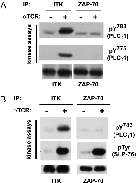
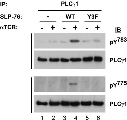
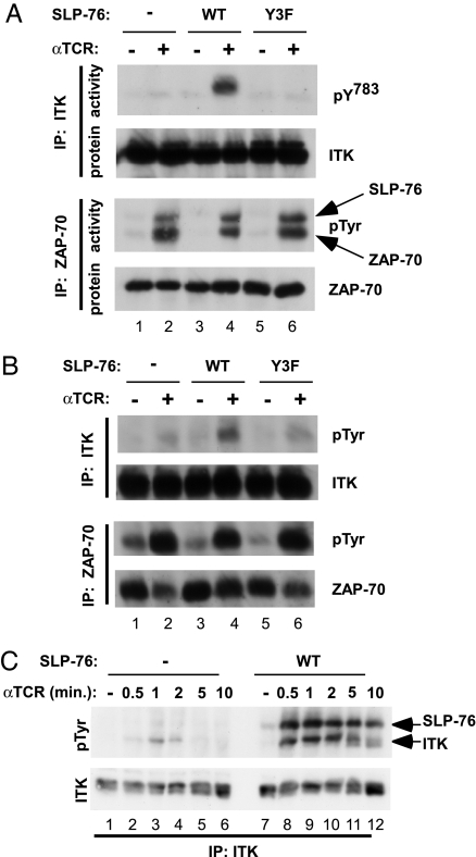
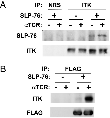
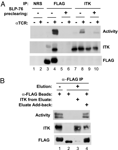
ITK (IL-2-inducible T cell kinase), a Tec family protein tyrosine kinase (PTK), is one of three PTKs required for T cell antigen receptor (TCR)-induced activation of phospholipase C-gamma1 (PLC-gamma1). Like Src and Abl family PTKs, ITK adopts an inactive, "closed" conformation, and its conversion to the active conformation is not well understood, nor have its direct substrates been identified. In a side-by-side comparison of ITK and ZAP-70 (zeta chain-associated protein kinase of 70 kDa), ITK efficiently phosphorylated Y(783) and Y(775) of PLC-gamma1, two phosphorylation sites that are critical for its activation, whereas ZAP-70 did not. SLP-76 (SH2-domain-containing leukocyte protein of 76 kDa), an adaptor required for TCR-induced activation of PLC-gamma1, was required for the phosphorylation of both PLC-gamma1 sites in intact cells. Furthermore, this event depended on the N-terminal tyrosines of SLP-76. Likewise, SLP-76, particularly its N-terminal tyrosines, was required for TCR-induced tyrosine phosphorylation and activation of ITK but was not required for the phosphorylation or activation of ZAP-70. Both ZAP-70 and ITK phosphorylated SLP-76 in vitro; thus, both PTKs are potential regulators of SLP-76, but only ITK is regulated by SLP-76. Upon TCR stimulation, a small fraction of ITK bound to SLP-76. This fraction, however, encompassed most of the catalytically active ITK. Catalytic activity was lost upon mild elution of ITK from the SLP-76-nucleated complex but was restored upon reconstitution of the complex. We propose that SLP-76 is required for ITK activation; furthermore, an ongoing physical interaction between SLP-76 and ITK is required to maintain ITK in an active conformation.


Conflict of interest statement

The authors declare no conflict of interest.

Figures

Fig. 1.
Fig. 1.
ITK, but not ZAP-70, efficiently phosphorylates both PLC-γ1 activation sites, Y783 and Y775, in vitro. J14 cells stably reconstituted with WT FLAG-tagged SLP-76 were mock-stimulated or stimulated for 1 min with anti-TCR and then lysed as described in Materials and Methods. (A) ITK or ZAP-70 immune complexes were prepared from the lysates of 5 × 10⁶ cells, and their catalytic activity was assayed, using a GST–PLCγ1SH2-SH2-SH3 fusion protein as a substrate. Kinase reaction products (top and middle blots) and immune complex beads (bottom blot) were separated by SDS/PAGE, and phospho-specific antibodies, anti-PLC-γ1 pY783 or anti-PLC-γ1 pY775, were used to detect phosphorylation of the GST–PLC-γ1 fusion protein. The beads were probed by Western blotting with anti-ITK and anti-ZAP-70 antibodies, as indicated. The data are representative of at least two independent experiments. (B) Conditions were the same as in A, except that the kinase substrate was GST–PLCγ1SH2-SH2-SH3 (top blot) or His-tagged SLP-76 (middle blot). Anti-pTyr (4G10) was used to detect phosphorylation of His-tagged SLP-76. The data are representative of at least two independent experiments. IP, immunoprecipitation.
Fig. 2.
Fig. 2.
SLP-76 and its N-terminal tyrosine phosphorylation sites are required for TCR-induced phosphorylation of PLC-γ1 at Y783 and Y775. J14-derived cell lines stably expressing the indicated FLAG-tagged alleles of SLP-76 were mock-stimulated or stimulated for 1 min with anti-TCR and lysed. Anti-PLCγ1 immune complexes prepared from lysates of 5 × 10⁶ cells were separated by SDS/PAGE and probed by Western blotting with phospho-specific antibodies (anti-PLCγ1 pY783 or anti-PLCγ1 pY775), as indicated. Each blot was then stripped and reprobed with anti-PLCγ1. The results are representative of at least three independent experiments. IB, immunoblot; IP, immunoprecipitation.
Fig. 3.
Fig. 3.
TCR-induced activation and tyrosine phosphorylation of ITK depend on SLP-76 and the N-terminal tyrosine phosphorylation sites of SLP-76. The indicated J14-derived cell lines were stimulated with anti-TCR for 1 min, and ITK or ZAP-70 was immunoprecipitated from the lysates, as indicated. (A) SLP-76 is required to activate ITK. Immune complexes were assayed for catalytic activity as in Fig. 1B, using GST–PLCγ1SH2-SH2-SH3 as a substrate for ITK (first blot) and His-tagged SLP-76 as a substrate for ZAP-70 (third blot). The electrophoretic mobility of the substrate (SLP-76) and enzyme (ZAP-70) are indicated by arrows on the right. In parallel, the immune complexes were probed for the presence of the relevant kinase (second and fourth blots). A representative result of at least three similar experiments is shown. (B) SLP-76 is required for TCR-induced phosphorylation of ITK. Immune complexes were probed with anti-phosphotyrosine and then stripped and reprobed with anti-ITK or anti-ZAP-70, respectively. (C) Time course of TCR-induced phosphorylation of ITK in the presence and absence of SLP-76. ITK was immunoprecipitated from the lysates of 10 × 106 cells, then probed with anti-phosphotyrosine, and then stripped and reprobed with anti-ITK. The bands corresponding to tyrosine-phosphorylated ITK and coimmunoprecipitating SLP-76 are indicated by arrows. This experiment was repeated three times with similar results. IP, immunoprecipitation.
Fig. 4.
Fig. 4.
TCR-inducible association of SLP-76 and ITK. J14 cells or J14 cells that were stably reconstituted with FLAG-tagged WT SLP-76 were mock-stimulated or stimulated with anti-TCR and then lysed as in Fig. 1. (A) Anti-ITK immune complexes were prepared from the lysates of 6 × 10⁶ cells and sequentially probed with anti-SLP-76 and then with anti-ITK. Normal rabbit serum (NRS) was used to control for nonspecific absorption to the beads. (B) Anti-FLAG immune complexes were prepared from the lysates of 15 × 106 cells and sequentially probed with anti-ITK and then with anti-FLAG. The data are representative of at least three independent experiments. IP, immunoprecipitation.
Fig. 5.
Fig. 5.
Catalytically active ITK is found in a complex with SLP-76. J14 cells stably reconstituted with FLAG-tagged WT SLP-76 were mock-stimulated or stimulated with anti-TCR and lysed as in Fig. 1. (A) SLP-76 was removed from the lysates by three rounds of immunoprecipitation with anti-FLAG (SLP-76 preclearing), or lysates were untreated (no preclearing). Subsequently, anti-FLAG immune complexes were prepared from the lysates of 15 × 106 cells, or anti-ITK immune complexes were prepared from the lysates of 5 × 10⁶ cells. Normal rabbit serum (NRS) was used to control for nonspecific absorption to the beads (lanes 1 and 2). Immune complexes were assayed for kinase activity, using GST–PLCγ1SH2-SH2-SH3 as a substrate (top blot). After the kinase assay, the immunoprecipitation beads were analyzed separately for the presence of ITK (middle blot) and FLAG-tagged SLP-76 (bottom blot). (B) Anti-FLAG immune complexes prepared from the lysates of 20 × 106 TCR-stimulated cells were left alone (lane 1) or were eluted with kinase reaction buffer supplemented with 0.2 M LiCl and then washed with kinase reaction buffer (lane 2). Eluates were desalted by dialysis against kinase reaction buffer and reimmunoprecipitated with anti-ITK (lane 3) or added back to the eluted anti-FLAG beads (lane 4). All beads were then washed before measuring in vitro kinase activity (top blot). After the reaction, the immunoprecipitation beads were analyzed for the presence of ITK (middle blot) and FLAG-tagged SLP-76 (bottom blot). IP, immunoprecipitation.

References

    1. Carpenter G, Ji Q. Exp Cell Res. 1999;253:15–24. - PubMed
    1. Rhee SG. Annu Rev Biochem. 2001;70:281–312. - PMC - PubMed
    1. Koretzky GA, Abtahian F, Silverman MA. Nat Rev Immunol. 2006;6:67–78. - PubMed
    1. Yablonski D, Weiss A. Adv Immunol. 2001;79:93–128. - PubMed
    1. Yablonski D, Kuhne MR, Kadlecek T, Weiss A. Science. 1998;281:413–416. - PubMed

Publication types

MeSH terms

Substances