Pure and mixed tubular carcinoma of the breast: mammographic and sonographic differential features
- PMID: 17420627
- PMCID: PMC2626773
- DOI: 10.3348/kjr.2007.8.2.103
Pure and mixed tubular carcinoma of the breast: mammographic and sonographic differential features
Abstract
Objective: We wanted to evaluate the mammographic and sonographic differential features between pure (PT) and mixed tubular carcinoma (MT) of the breast.
Materials and methods: Between January 1998 and May 2004, 17 PTs and 14 MTs were pathologically confirmed at our institution. The preoperative mammography (n = 26) and sonography (n = 28) were analyzed by three radiologists according to BI-RADS.
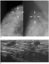
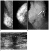
Results: On mammography, a mass was not detected in eight patients with PT and in one patient with MT (57% vs. 8%, respectively, p = 0.021), which was statistically different. The other findings on mammography and sonography showed no statistical differences between the PT and MT, although the numerical values were different. When the lesions were detected mammographically, an irregularly shaped mass with a spiculated margin was more frequently found in the MT than in the PT (100% vs. 83%, respectively, p = 0.353). On sonography, all 28 patients presented with a mass and most lesions showed as not being circumscribed, hypoechoic masses with an echogenic halo. Surrounding tissue changes and posterior shadowing were more frequently found in the MT than in the PT (75% vs. 50%, respectively, p = 0.253, 58% vs. 19%, respectively, p = 1.000). An oval shaped mass was more frequently found in the PT than in the MT (44% vs. 25%, respectively; p = 0.434).
Conclusion: PT and MT cannot be precisely differentiated on mammography and sonography. However, the absence of a mass on mammography or the presence of an oval shaped mass would favor the diagnosis of PT. An irregularly shaped mass with surrounding tissue change and posterior shadowing on sonography would favor the diagnosis of MT and also a less favorable prognosis.
Figures


References
-
- Tavassoli FA, Devilee P. Pathology and genetics. In: Jaffe ES, Harris NL, Stein H, editors. World Health Organization classification of tumours: tumours of the breast and female genital organs. Lyon: IARC Press; 2003. pp. 26–28.
-
- Rosen PP. Rosen's breast pathology. 2nd ed. Philadelphia: Lippincott-Williams & Wilkins; 2001. pp. 365–380.
-
- Deos PH, Norris HJ. Well-differentiated (tubular) carcinoma of the breast. A clinicopathologic study of 145 pure and mixed cases. Am J Clin Pathol. 1982;78:1–7. - PubMed
-
- Leibman AJ, Lewis M, Kruse B. Tubular carcinoma of the breast: mammographic appearance. AJR Am J Roentgenol. 1993;160:263–265. - PubMed
-
- Sheppard DG, Whitman GJ, Huynh PT, Sahin AA, Fornage BD, Stelling CB. Tubular carcinoma of the breast: mammographic and sonographic features. AJR Am J Roentgenol. 2000;174:253–257. - PubMed
MeSH terms
LinkOut - more resources
Full Text Sources
Medical

