Review
. 2007 Jul;236(7):1841-64.
doi: 10.1002/dvdy.21138.

How to innervate a simple gut: familiar themes and unique aspects in the formation of the insect enteric nervous system

Affiliations
Review

How to innervate a simple gut: familiar themes and unique aspects in the formation of the insect enteric nervous system

Philip F Copenhaver. Dev Dyn. 2007 Jul.

Abstract

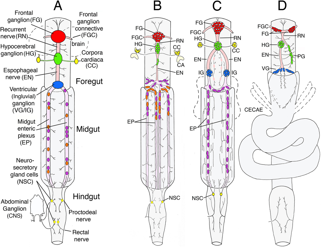
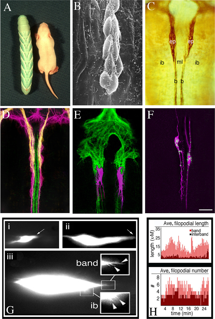
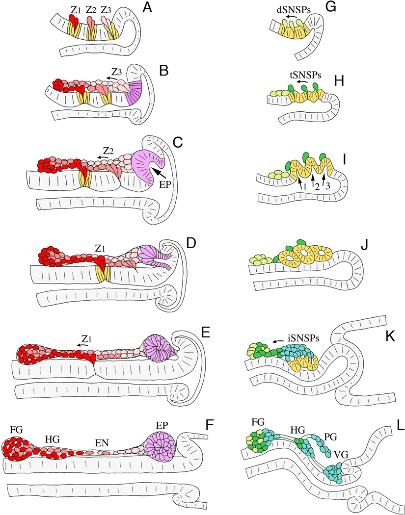
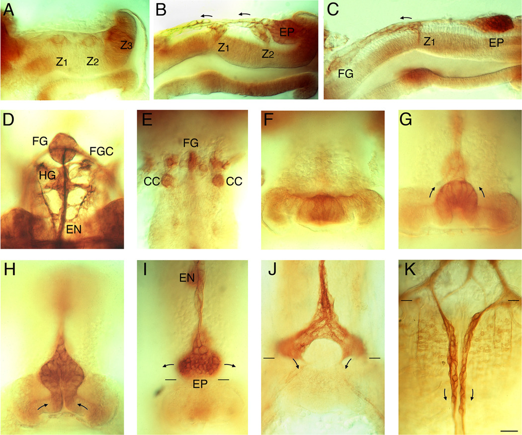
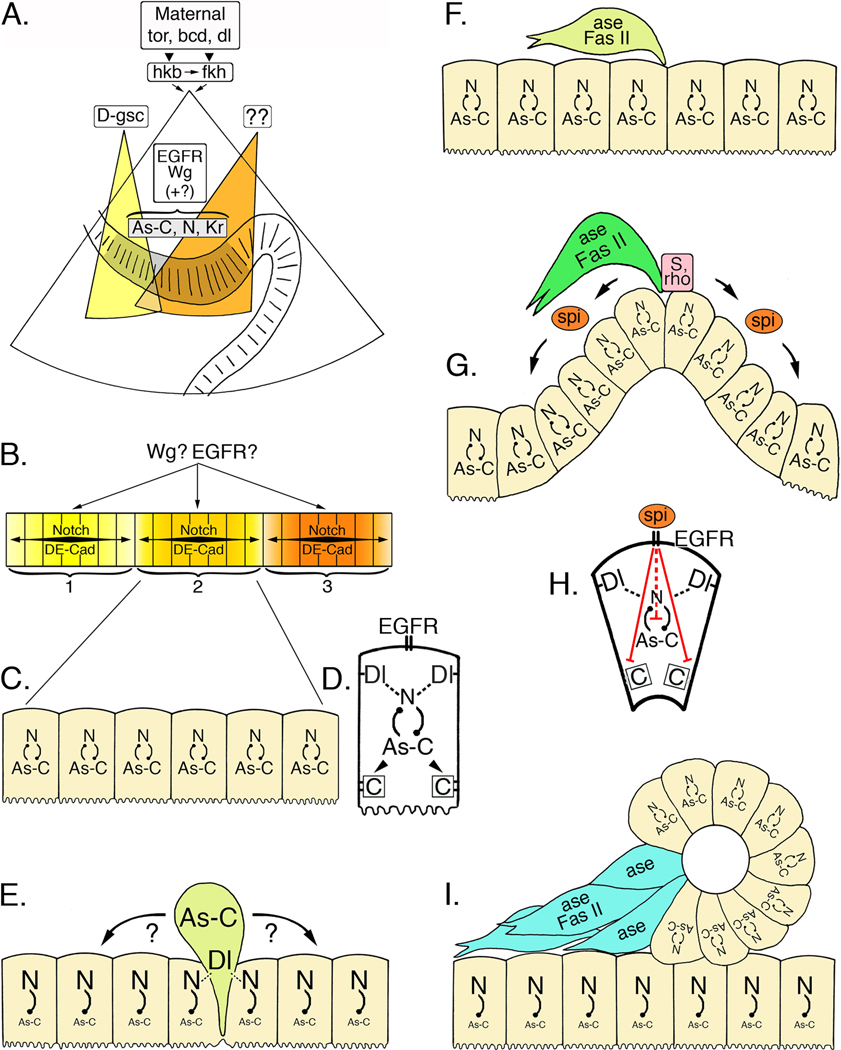
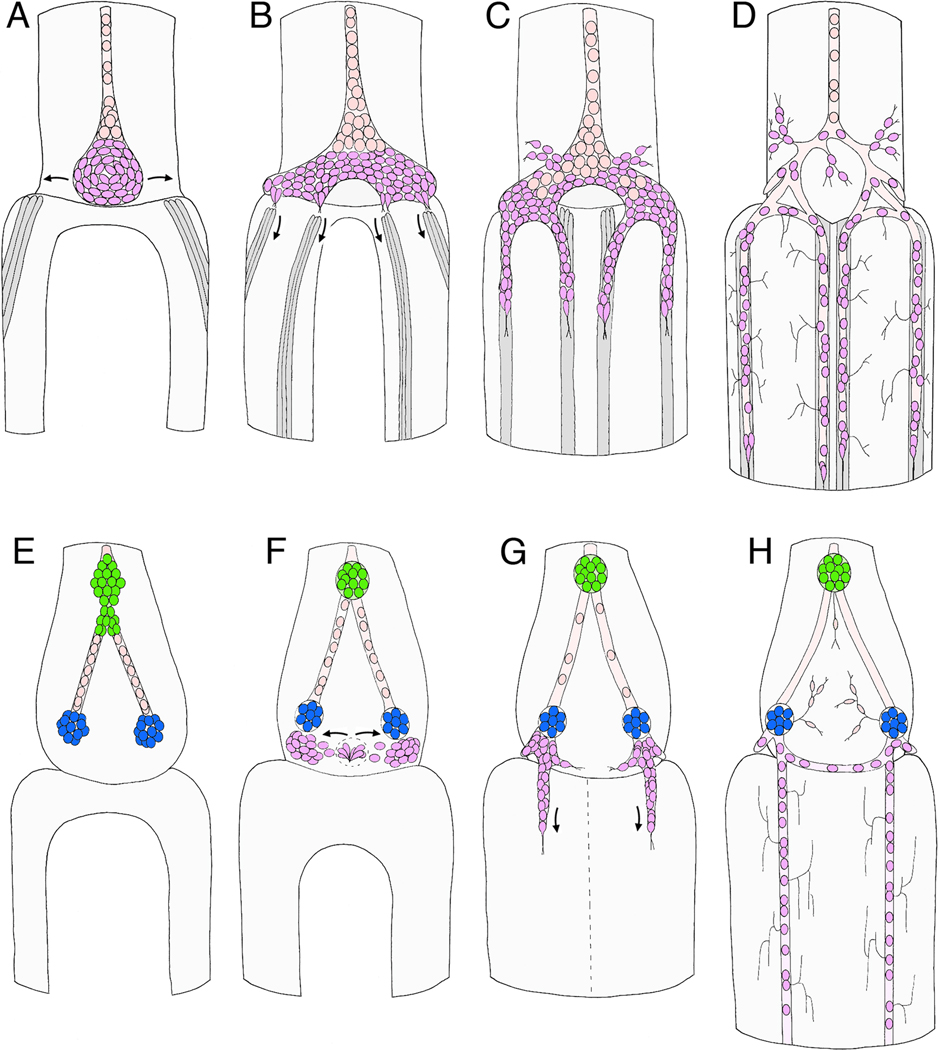
Like the vertebrate enteric nervous system (ENS), the insect ENS consists of interconnected ganglia and nerve plexuses that control gut motility. However, the insect ENS lies superficially on the gut musculature, and its component cells can be individually imaged and manipulated within cultured embryos. Enteric neurons and glial precursors arise via epithelial-to-mesenchymal transitions that resemble the generation of neural crest cells and sensory placodes in vertebrates; most cells then migrate extensive distances before differentiating. A balance of proneural and neurogenic genes regulates the morphogenetic programs that produce distinct structures within the insect ENS. In vivo studies have also begun to decipher the mechanisms by which enteric neurons integrate multiple guidance cues to select their pathways. Despite important differences between the ENS of vertebrates and invertebrates, common features in their programs of neurogenesis, migration, and differentiation suggest that these relatively simple preparations may provide insights into similar developmental processes in more complex systems.


Figures

Figure 1
Figure 1. Schematic drawings of the ENS in different insect species
A. Generalized organization of ganglia and nerves found in the insect ENS. The primary foregut ganglion is the frontal ganglion (FG; red), connected to the overlying brain (dotted outline) by paired frontal ganglion connectives (FGC). Several nerve branches extend anteriorly onto the pharynx, while the recurrent nerve (RN) extends posteriorly to the hypocerebral ganglion (HG; green), situated below the brain. The HG is also usually connected to the paired corpora cardiaca (CC), the primary neurosecretory organs of the brain. From the HG, the esophageal nerve (EN) extends to the ventricular ganglion (VG; blue; also called the ingluvial ganglion, or IG). The foregut ganglia also give rise to a diffuse plexus of nerves that innervate the musculature and may include peripheral stretch receptors. From the ventricular ganglion, a branching nerve plexus (the midgut enteric plexus, EP) extends along the superficial musculature of the midgut; typically, this plexus contains a distributed set of enteric neurons within its major branches. The hindgut is innervated by branches of the proctodeal and rectal nerves that originate in the terminal abdominal ganglion of the ventral nerve cord (CNS); branches of the proctodeal nerve also extend onto the posterior midgut. Several peripheral neurosecretory cells are often found within these nerves (yellow). B. Diagram of the ENS in the tobacco hornworm Manduca sexta (larval stage). The FG is closely apposed to a diminutive HG, but no VG forms in this species. The esophageal nerve connects with the enteric plexus that spans the foregut-midgut boundary and contains dispersed populations of neurons. Several distinct neuronal phenotypes are intermingled within the anterior portion of the midgut nerves (orange and purple cells). C. Diagram of the ENS in the grasshopper Schistocerca americana (after Ganfornina et al., 1996). Posterior to the HG, two esophageal nerves extend to paired IG near the foregut-midgut boundary; nerves from these ganglia connect with the midgut enteric plexus, which contains distributed neurons throughout its length in this species. D. Diagram of the ENS in the fruit fly Drosophila melanogaster (after Skaer, 1993 and Hartenstein et al., 1994). The FG consists of an asymmetric pair of hemi-ganglia on either side of the foregut. Branches of the recurrent nerve extend to the HG on the left and to the paraesophageal ganglion (PG) on the right. From the HG, the esophageal nerve extends to a single VG, consisting of a small number of neurons near the foregut-midgut boundary. Two sets of nerves extend from the VG onto the anterior portion of the midgut, but these nerves do not contain enteric neurons.
Figure 2
Figure 2. Manduca as an experimental model offers the advantages of simplicity and accessibility, permitting a variety of manipulations to be performed in the intact ENS
A. Photograph of a fully grown Manduca larva (fifth instar) and a juvenile mouse (courtesy of Dr. Rita Balice-Gordon). B. Scanning electron micrograph of EP cells that are migrating on the surface of the gut musculature. C. Whole-mount preparation of a Manduca embryo immunostained with an antibody against the cell adhesion receptor Fas II (at 58% of development; 1% of development = 1 hr). Both the EP cells (ep) and their muscle band pathways (b) express Fas II at this stage of development. EP cells migrate exclusively on the midgut bands but not onto adjacent interband muscles (ib), nor across the midline interband cells (ml). Only the dorsal pair of eight midgut bands is shown. D. Whole-mount preparation of the embryonic ENS (at 65% of development) immunostained with anti-MsEphrin (magenta; to label the EP ells and their processes); anti-GPI-Fas II (yellow; to label the glial cells ensheathing the midgut EP cells), and anti-Neuroglian (green; to label the muscle band pathways). E. Whole-mount preparation of the embryonic ENS (at 58% of development) in which two migratory EP cells were injected with DiI (magenta) prior to immunostaining the preparation with anti-TM-Fas II (green). As in panel C, the muscle band pathways are also stained by anti-Fas II antibodies. Each neuron extends an array of filopodial processes in advance of its cell body; filopodia that extend along the muscle band pathways are longer and are often become incorporated into the leading process, while filopodia that extend onto the adjacent interband muscles remain shorter and usually are rapidly retracted. F. Embryo in which two EP cells were injected prior to migration onset (at 50% of development) with mRNA encoding monomeric DsRed and Alexa Fluor 488 hydrazide (green). After 20 hr in culture, the preparation was immunostained with anti-DsRed antibodies (magenta). G. Single frames taken from QuickTime movies of EP cells that were injected with Alexa Fluor-488 dextran and monitored by time-lapse imaging. Panel “i” shows an EP cell migrating along one of the midgut muscle bands (the band is not labeled); arrow indicates the position of the leading process (see Supplementary movie #S1). Panel “ii” shows an EP cell transitioning from migration to axonal outgrowth (see Supplementary movie #S2). Arrow indicates the position of the leading process that will form the axon. Panel “iii” shows a higher magnification image of a migrating EP cell to visualize the filopodia associated with its leading process (see Supplementary movie #S3). Boxed insets indicate filopodia extending onto its muscle band pathway (band) and onto adjacent interband musculature (ib) that are quantified in panel H. H. Filopodial dynamics of a migrating EP cell over the course of 30 min (data collected from Supplementary movie #S3); upper panel indicates the average length of filopodia extending along the band pathway (red histograms) or adjacent interband musculature (shaded histograms). Lower panel shows the average number of filopodia on the band versus interband regions. Scale = 0.75 cm in A; 10 µm in B; 40 µm in C–D; 20 µm in E–F. The average somal length of the migrating EP cells in panel G is ~15 µm.
Figure 3
Figure 3. Schematic illustration of neurogenesis in Manduca (A–F; after Copenhaver and Taghert, 1991) and Drosophila (G–L; after Hartenstein, 1997)
Each panel represents a sagittal view of the foregut midline; anterior is to the left, dorsal is to the top. A. At ~24% of development in Manduca, three neurogenic zones have formed in the dorsal foregut epithelium (Z1, Z2, & Z3), from which a series of individual precursors will delaminate; each precursor cell divides once or twice after emerging. B. By 28% of development, streams of zone-derived cells have begun to migrate anteriorly along the foregut, while all of the remaining zone 3 cells delaminate. The epithelium surrounding the original position of zone 3 subsequently differentiates into a distinct placode (purple). C. By 33% of development, migrating zone cells have begun to form the frontal ganglion anteriorly, while all of the remaining zone 2 cells delaminate. The EP cell placode has also begun to invaginate. D. At 36% of development, zone-derived cells continue to migrate along the pathways that will form the recurrent and esophageal nerves, while the EP cell placode invaginates. E. By 39% of development, the last of the zone 1 cells delaminate, and the EP cell placode has invaginated onto the foregut surface. F. By 42% of development, the frontal and hypocerebral ganglia have formed; subsets of the residual zone cells will proliferate to form glial populations that ensheath the nerves and ganglia of the ENS. At this stage, the invaginated EP cells form a condensed packet of post-mitotic neurons adjacent to the foregut-midgut boundary. G. In Drosophila at stage 10 (~24% of development), three neurogenic centers form within the ENS anlage (yellow) and give rise to a set of delaminating precursors (dSNSPs; light green). H. At stage 11 (~30% of development), the dSNSPs have migrated anteriorly, where they will help form the frontal ganglion and its nerves. A second wave of precursors (tSNSPs, dark green) delaminates from the neurogenic centers, marking the positions where three invaginations will form. I. By stage 12 (~34% of development), the three invaginations form distinct pouches (1, 2, & 3) that protrude onto the foregut surface. J. By stage 13 (~38% of development), the three pouches have pinched off to form a set of neurogenic vesicles, while the tSNSPs migrate anteriorly to help form the frontal and hypocerebral ganglia. K. By stage 14 (~42% of development), invaginated cells from the three vesicles begin to dissociate (iSNSPs) and migrate anteriorly. L. By stage 16 (~60% of development), the SNSPs have assembled into the enteric ganglia of the foregut: frontal ganglion (FG), hypocerebral ganglion (HG), paraesophageal ganglion (PG), and ventricular ganglion (VG). Processes from these neurons also pioneer the interganglionic nerves.
Figure 4
Figure 4. Photomicrographs of the developing ENS in Manduca (immunostained with anti-Fas II antibodies; after Copenhaver and Taghert, 1990, 1991)
A–C. Lateral views of the foregut at 24%, 33%, and 39% of development. Z1, Z2, and Z3 indicate the three neurogenic zones of the foregut; EP = the invaginating placode giving rise to the migratory neurons that populate the midgut. D. Dorsal view of the frontal ganglion (FG) and hypocerebral ganglion (HG) at ~60% of development; the frontal ganglion connectives to the brain (FGC) and posterior esophageal nerve (EN) are also visible. E Dorsal view of a younger embryo (~35% of development) shows the emergence of cells that will migrate off the foregut to form the intrinsic neurons of the corpora cardiaca (CC). F–H: dorsal views of the posterior lip of the foregut at 30%, 34%, and 38% of development, showing the invagination of the EP cell placode. I. By 42% of development, the EP cells have invaginated to form a packet of post-mitotic neurons adjacent to the foregut-midgut boundary (paired black lines). Anteriorly, the EP cell packet is in continuity with the residual zone 3 cells and the developing esophageal nerve (EN). Arrows indicate the directions the EP cells will subsequently follow as they spread bilaterally around the foregut. J. By 55% of development, the EP cells have spread almost completely around the foregut and have begun to align with eight longitudinal muscle bands that form on the midgut as it closes dorsally. Arrows indicate the direction that subsets of EP cells will follow once migration onto the midgut commences (the dorsal pair of muscle bands can be faintly seen below the arrows). K. By 58%, subsets of EP cells have begun to migrate rapidly along the muscle band pathways on the midgut; only the dorsal pair of eight parallel bands are shown. Scale = 40 µm.
Figure 5
Figure 5. A cascade of regulatory genes controls neurogenesis in the ENS of Drosophila
A. Maternally expressed torso (tor), bicoid (bcd), and dorsal (dl) control the expression of huckebein (hkb) and fork head (fkh) in the invaginating stomodeum (after Hartenstein, 1997). The homeobox gene D-goosecoid (D-gsc; yellow field) is required for the differentiation of the anterior stomodeum, including the anterior neurogenic center (zone 1) of the ENS. Other patterning genes (as yet unidentified; orange field) may specify the formation of the more posterior zones. As the stomodeum invaginates, the ENS anlage (grey shaded epithelium) becomes morphologically distinguishable and begins to express a combination of proneural genes in the Achaete-scute Complex (As-C), neurogenic genes (including Notch; N), and several other transcription factors, including Krüppel (Kr). Both Wingless (Wg) and EGFR signaling (plus other identified regulatory genes) play essential but poorly defined roles in this initial phase of ENS development. B. Wg and EGFR signaling may also help delineate the three neurogenic centers (1, 2, & 3) that subsequently form within the ENS anlage, possibly by limiting the range of Notch signaling within each zone and by modulating cell adhesive interactions mediated by Drosophila E-Cadherin (DE-Cad). C. All of the cells within each zone initially express intermediate levels of both proneural genes (As-C) and neurogenic genes (N). D. Enlarged view of a single zone cell at this stage. Notch (N)-Delta (Dl) interactions between adjacent cells are regulated in part by inhibitory feedback with the proneural genes (As-C); proneural genes may also regulate DE-Cadherin expression (boxed C). E. During the sequential delamination of individual precursors, lateral inhibition by the neurogenic genes promotes enhanced expression of the proneural genes (As-C) in a single zone cell, which then emerges from the foregut epithelium (light green cell represents a dSNSP). Cadherin-mediated adhesive interactions must also be down-regulated at this time. Concurrently, elevated levels of Notch signaling in the remaining zone cells help maintain their epithelial organization. F. Once delaminated, the precursor cell expresses the proneural gene Asense (ase), which may restrict further mitotic divisions, and the cell adhesion receptor Fas II, which may promote directed migration. The remaining zone cells re-acquire a balanced expression of both proneural genes and neurogenic genes. G. A second cycle of delamination gives rise to another set of precursors that emerge onto the foregut (dark green cell represents a tSNSP). As these precursors emerge, elevated levels of Star (S) and Rhomboid (rho) result in the localized release of the EGFR ligand, Spitz (spi), which in turn promotes the invagination of the remaining zone cells. H. Enlarged view of a single invaginating cell; EGFR signaling induced by Spitz interrupts the normal inhibitory feedback between neurogenic and proneural genes, permitting their continued co-expression. EGFR signaling also down-regulates DE-Cadherin-mediated adhesion, thereby promoting the morphological reorganization of the invaginating cells. I. Invagination of the neurogenic zone produces a discrete epithelial vesicle (see Fig. 3J), in which all of the cells continue to express both proneural genes (maintaining their potential to become neurons) and neurogenic genes (which help maintain their epithelial organization). By contrast, proneural gene expression is inhibited in the underlying epithelial layer. Individual cells from the vesicle then down-regulate neurogenic gene expression and disperse, while they upregulate Asense and Fas II (blue cells represent iSNSPs).
Figure 6
Figure 6. Schematic illustrations of the sequence of migration that forms the midgut enteric plexus in the insect ENS
A–D represents Manduca (after Copenhaver and Taghert, 1989b; Copenhaver, 1993); E–H represents Schistocerca (after Ganfornina et al., 1996). All panels show dorsal views of the developing ENS at the foregut-midgut boundary. A. At 40% of development, the EP cells have invaginated onto the dorsal foregut surface and begin to spread bilaterally around the foregut-midgut boundary. Concurrently, subsets of longitudinal muscles on the midgut (dark grey cells) begin to coalesce as dorsal closure of the midgut proceeds. Anteriorly, the EP cell packet is in continuity with the residual zone 3 cells that help form the esophageal nerve; subsets of these zone-derived cells will subsequently proliferate to form glial cells that ensheath the enteric plexus. B. By 55% of development, the EP cells have almost completely surrounded the foregut, and subsets of the cells have begun to align with each of the eight midgut muscle bands (only the dorsal four are shown). C. By 58% of development, the EP cells have begun to migrate posteriorly along the muscle bands on the midgut; a small number of neurons also migrate laterally onto radial muscles of the foregut (foregut muscles not shown). D. By 80%, the EP cells have completed their migration, forming the enteric plexus that spans the foregut-midgut boundary; they have also extended axons along the posterior midgut (not shown) and short terminal branches onto the adjacent interband musculature. Glial cells (pink) derived from the residual zone 3 cells have also migrated over the major branches of the enteric plexus to ensheath them. E. By 40% in the Grasshopper ENS, the neurogenic zones of the foregut have given rise to the cells of frontal ganglion (not shown) and hypocerebral ganglion (green), while posteriorly, cells derived from the third neurogenic zone have migrated bilaterally to form the incipient ingluvial ganglia (blue). F. By ~50% of development, a second wave of neurogenesis from the vicinity of zone 3 has begun to produce a new population of ingressing cells (purple); these cells then migrate bilaterally and aggregate adjacent to the ingluvial ganglia. G. By ~60% of development, four streams of cells have begun to migrate posteriorly from the ingluvial ganglia onto the midgut (only the dorsal pair is shown). Unlike Manduca, distinct muscle bands on the midgut have not been detected in grasshopper. H. By 80% of development, the migratory populations of neurons have become distributed along the entire length of the midgut and have extended terminal branches onto the adjacent musculature. An additional set of cells derived from the neurogenic zones (presumably sensory neurons) also contributes to an extensive foregut plexus (not detected in Manduca).

Similar articles

Cited by

References

    1. Airaksinen MS, Holm L, Hatinen T. Evolution of the GDNF family ligands and receptors. Brain Behav Evol. 2006;68:181–190. - PubMed
    1. Alberti S, Krause SM, Kretz O, Philippar U, Lemberger T, Casanova E, Wiebel FF, Schwarz H, Frotscher M, Schutz G, Nordheim A. Neuronal migration in the murine rostral migratory stream requires serum response factor. Proc Natl Acad Sci U S A. 2005;102:6148–6153. - PMC - PubMed
    1. Anderson DJ. Stem cells and transcription factors in the development of the mammalian neural crest. Faseb J. 1994;8:707–713. - PubMed
    1. Anderson DJ. Lineages and transcription factors in the specification of vertebrate primary sensory neurons. Curr Opin Neurobiol. 1999;9:517–524. - PubMed
    1. Anderson RB, Turner KN, Nikonenko AG, Hemperly J, Schachner M, Young HM. The cell adhesion molecule L1 is required for chain migration of neural crest cells in the developing mouse gut. Gastroenterology. 2006;130:1221–1232. - PubMed

Publication types

MeSH terms

LinkOut - more resources