Autoradiographic and immunohistochemical study on the proliferative kinetics of intestinal metaplasia
- PMID: 1742257
- PMCID: PMC4535013
- DOI: 10.3904/kjim.1991.6.1.8
Autoradiographic and immunohistochemical study on the proliferative kinetics of intestinal metaplasia
Abstract
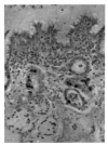
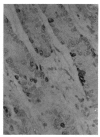
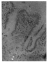
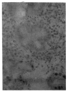
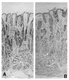
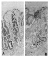
In order to elucidate the proliferative behavior of the intestinal metaplasia around gastric cancer, the authors used both in vitro tritiated thymidine (3H-thymidine) autoradiography and in vivo bromodeoxyuridine (BrdUrd) immunohistochemistry for labeling the proliferative cells of the normal pyloric glands and metaplastic gastric glands. The results of the methods were comparable: The labeling pattern and the rate of labeling were very similar. In the normal pyloric mucosa, the labeled cells were confined to the isthmus region, indicating that pyloric glandular cells are normally renewed from the isthmus region. On the other hand, a zone of the labeled cells was found in the lower half of the intestinalized mucosa, indicating that cell proliferation took place deep in the mucosa, just like the case of normal intestinal glands. The labeling indices of the pyloric mucosa were 19.4% by autoradiography and 18.0% by immunohistochemistry, and that of the intestinalized gastric glands were 25.2% by autoradiography and 24.2% by immunohistochemistry. In conclusion, both 3H-thymidine autoradiography and BrdUrd immunohistochemistry showed that the proliferative kinetics of the intestinalized gastric glands was similar to that of the normal intestinal glands rather than the pyloric glands, i.e. a lower level of proliferative zone and higher labeling index were present.
Figures

References
-
- Korn ER. Intestinal metaplasia of the gastric mucosa. Am J Gastroenterol. 1973;60:270. - PubMed
-
- Yoon CM, Rew JS, Park KS. Subtypes of intestinal metaplasia in endoscopic biopsies of various gastric diseases. Kor J Gastroenterol. 1987;19:13.
-
- Ming SC, Goldman H, Freiman DG. Intestinal metaplasia and histogenesis of carcinoma in human stomach. Cancer. 1967;20:1418. - PubMed
-
- Jarvi O, Lauren P. On the role of heterotopias of the intestinal epithelium in the pathogenesis of gastric cancer. Acta Path MicrobiolScand. 1965;64:31. - PubMed
-
- Kawachi T, Kurisu M, Numanyu N, Sasajima K, Sano T, Sugimura T. Precancerous changes in the stomach. Cancer Res. 1976;36:2673. - PubMed
MeSH terms
LinkOut - more resources
Full Text Sources
