. 2007 May;5(5):e97.
doi: 10.1371/journal.pbio.0050097.

Structural and chemical profiling of the human cytosolic sulfotransferases

Affiliations

Structural and chemical profiling of the human cytosolic sulfotransferases

Abdellah Allali-Hassani et al. PLoS Biol. 2007 May.

Erratum in

  • PLoS Biol. 2007 Jun;5(6):e165. Thonton, Janet [corrected to Thornton, Janet]

Abstract

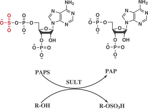
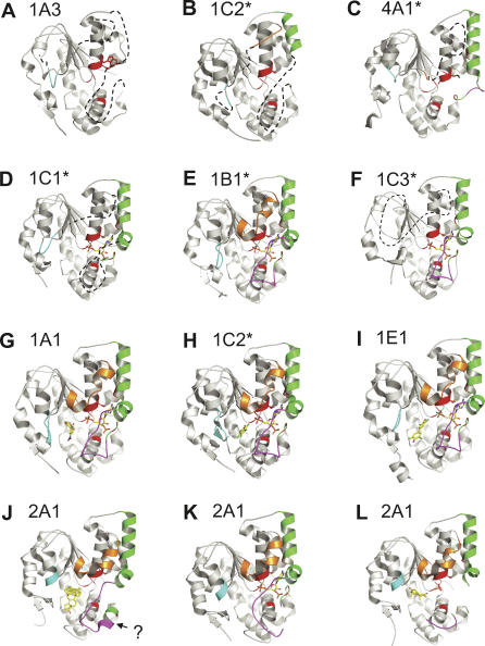
The human cytosolic sulfotransfases (hSULTs) comprise a family of 12 phase II enzymes involved in the metabolism of drugs and hormones, the bioactivation of carcinogens, and the detoxification of xenobiotics. Knowledge of the structural and mechanistic basis of substrate specificity and activity is crucial for understanding steroid and hormone metabolism, drug sensitivity, pharmacogenomics, and response to environmental toxins. We have determined the crystal structures of five hSULTs for which structural information was lacking, and screened nine of the 12 hSULTs for binding and activity toward a panel of potential substrates and inhibitors, revealing unique "chemical fingerprints" for each protein. The family-wide analysis of the screening and structural data provides a comprehensive, high-level view of the determinants of substrate binding, the mechanisms of inhibition by substrates and environmental toxins, and the functions of the orphan family members SULT1C3 and SULT4A1. Evidence is provided for structural "priming" of the enzyme active site by cofactor binding, which influences the spectrum of small molecules that can bind to each enzyme. The data help explain substrate promiscuity in this family and, at the same time, reveal new similarities between hSULT family members that were previously unrecognized by sequence or structure comparison alone.


Conflict of interest statement

Competing interests. The authors have declared that no competing interests exist.

Figures

Figure 1
Figure 1. Schematic of the Reaction Catalyzed by SULTs
Figure 2
Figure 2. Representative Structures for Each of the hSULTs Grouped According to Similar Global Structural Features
Apo proteins with considerable disorder in several loops: (A) SULT1A3 (1CJM), (B) SULT1C2 (2AD1), and (C) SULT4A1 (1ZD1). Structures with increased order due to binding of PAP (green helices, purple loop): (D) SULT1C1 (2ETG), (E) SULT1B1 (1XV1), and (F) SULT1C3 (2H8K). Ternary complexes display further ordering of substrate binding loops: (G) SULT1A1 with PAP and p-nitrophenol (1LS6), (H) SULT1C2 with PAP and PCP (2GWH), and (I) 1E1 with PAP and 3,5,3′,5′-tetrachloro-biphenyl-4,4'-diol (1G3M). Structures with unusual features that likely reflect catalytically unproductive proteins: (J) SULT2A1 bound to DHEA, but without PAP (1J99); compare to (K) structure of the same protein with PAP (1EFH), and (L) with androsterone (1OV4). The structures we solved are labelled with an asterisk. The question mark (?) indicates a helix formation, which leads to a non-productive conformation. The Protein Data Bank code for each structure is shown in parentheses. The proteins are represented by a ribbon model; PAP and substrate are shown as stick models coloured per element (carbon in yellow, oxygen in red, nitrogen in blue, and phosphate in magenta). The loops are coloured as discussed in the text (gold, cyan, green, and purple).
Figure 3
Figure 3. Sequence Alignment of hSULTs Showing Structural Features with Colour Coding Corresponding to That of the Structures in Figure 2
Figure 4
Figure 4. Thermo-denaturation/Aggregation Profile of SULT2A1
The intensity of scattered light is plotted as a function of temperature (T) for SULT2A1 in the absence of any ligand (filled diamond), and in the presence of AMP-PNP (filled square), lithocholic acid (open triangle), PAP (filled triangle), and both PAP and dehydroisoandrosterone 3-sulfate (open circle). The inflection point of the denaturation curve is taken as T agg as reported in Table 1.
Figure 5
Figure 5. Inhibitory Effect of Isoprenaline, PLP, Quercetin, DBHD, and PCP on Sulfotransferase Activity of SULT1B1 with 1-Naphthol as Substrate
Sulfotransferase activity of SULT1B1 at different concentrations of each compound (5 to 312 μM) has been assayed as described in Materials and Methods. Isoprenaline is indicated by a filled diamond, PLP by an open circle, quercetin by a filled square, DBHD by a filled triangle, and PCP by a filled circle.
Figure 6
Figure 6. The Superimposition of the Active Sites of the SULT1E1-PAP-17β-estradiol Complex (1AQU) and the SULT1C2-PAP-PCP Complex (2GWH)
The SULT1E1-PAP-17β-estradiol complex is shown in yellow, and the SULT1C2-PAP-PCP complex is shown in grey. The PCP (chlorine atoms in green) and 17β-estradiol (EST) molecules are highlighted along with the catalytic His115 and PAP. See text for discussion.
Figure 7
Figure 7. SULT4A1 Does Not Bind PAP
(A) Thermal aggregation temperatures for SULT1B1 (filled circle), SULT1C3 (filled triangle), SULT1C1 (filled diamond), SULT1C2 (filled square), and SULT4A1 (open circle) as a function of PAP concentration. PAP provides no stabilizing effect to SULT4A1. (B) Superimposition of the PAP binding site of SULT1C1 (green) and SULT4A1 (yellow). A conserved Trp residue (W53 of SULT1C1; green ring in the middle of figure, indicated by a red arrow) normally forms π-π stacking interactions with the adenine ring of PAP (blue) and hydrogen bonds to a neighbouring conserved Thr (T228 in SULT1C1). SULT4A1 is the only SULT to have a Leu instead of Trp at this position. The Leu residue cannot make stabilizing interactions with Thr or the adenine ring, and its position would cause a severe steric clash with PAP and PAPS.
Figure 8
Figure 8. Average Linkage Hierarchical Clustering of SULTs
Clustering is shown according to (A) global sequence similarity, (B) local sequence of the substrate binding site, (C) local structure of the substrate binding site, (D) catalytic activity profiles, (E) small molecule binding profiles in the presence of PAP, and (F) small molecule binding profiles in the absence of PAP. The value under each clustering (c) is the correlation between the original data matrix and the cophenetic matrix—a matrix whose elements represent the height at which these elements first meet in the tree. The higher this correlation, the more accurate the tree represents the original data. See Materials and Methods for further details.
Figure 9
Figure 9. Binding Site Comparison of SULT1A1 Bound to PAP and p-Nitrophenol (1LS6) and SULT1A3 Bound to PAP and Dopamine (2A3R)
The side chains of the eight binding-site residues that differ between these two proteins are shown in yellow (SULT1A1) and red (SULT1A3).

References

    1. Glatt H. Sulfotransferases in the bioactivation of xenobiotics. Chem Biol Interact. 2000;129:141–170. - PubMed
    1. Blanchard RL, Freimuth RR, Buck J, Weinshilboum RM, Coughtrie MW. A proposed nomenclature system for the cytosolic sulfotransferase (SULT) superfamily. Pharmacogenetics. 2004;14:199–211. - PubMed
    1. Freimuth RR, Wiepert M, Chute CG, Wieben ED, Weinshilboum RM. Human cytosolic sulfotransferase database mining: Identification of seven novel genes and pseudogenes. Pharmacogenomics J. 2004;4:54–65. - PubMed
    1. Hildebrandt MA, Salavaggione OE, Martin YN, Flynn HC, Jalal S, et al. Human SULT1A3 pharmacogenetics: gene duplication and functional genomic studies. Biochem Biophys Res Commun. 2004;321:870–878. - PubMed
    1. Falany CN, Xie X, Wang J, Ferrer J, Falany JL. Molecular cloning and expression of novel sulphotransferase-like cDNAs from human and rat brain. Biochem J. 2000;346(Pt 3):857–864. - PMC - PubMed

Publication types

Associated data