. 2007 May;121(1):71-81.
doi: 10.1111/j.1365-2567.2006.02539.x.

Peroxisome proliferator-activated receptor-gamma agonists inhibit respiratory syncytial virus-induced expression of intercellular adhesion molecule-1 in human lung epithelial cells

Affiliations

Peroxisome proliferator-activated receptor-gamma agonists inhibit respiratory syncytial virus-induced expression of intercellular adhesion molecule-1 in human lung epithelial cells

Ralf Arnold et al. Immunology. 2007 May.

Abstract

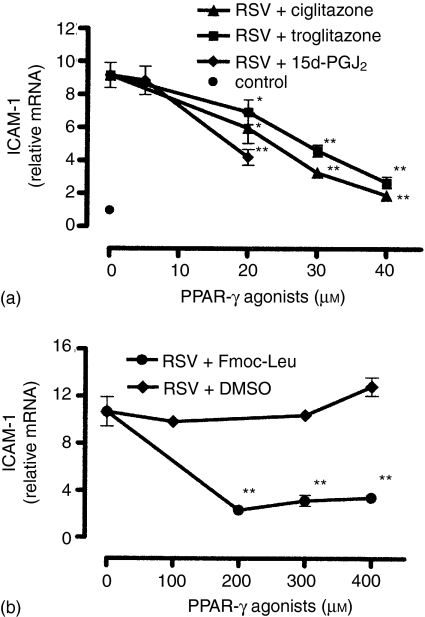
Respiratory syncytial virus (RSV) is the major causative agent of severe lower respiratory tract disease and death in infants worldwide. The epithelial cells of the airways are the target cells for RSV infection and the site of the majority of the inflammation associated with the disease. However, despite five decades of intensive RSV research there exist neither an effective active vaccine nor a promising antiviral and anti-inflammatory therapy. Recently, peroxisome proliferator-activated receptor-gamma (PPAR-gamma), a member of the nuclear hormone receptor superfamily, has been shown to possess anti-inflammatory properties. Therefore, we hypothesized whether the detrimental increase of intercellular adhesion molecule-1 (ICAM-1) on RSV-infected lung epithelial cells (A549 and primary normal human bronchial epithelial cells (NHBE)) might be modulated by natural and synthetic PPAR-gamma agonists (15d-PGJ2, ciglitazone, troglitazone, Fmoc-Leu). Our data show that all PPAR-gamma agonists under study significantly down-regulated the RSV-induced expression of ICAM-1 on A549- and NHBE cells in a dose-dependent manner resulting in a reduced beta2 integrin-mediated adhesion of monocytic effector cells (U937) to RSV-infected A549 cell monolayers. In contrast, the PPAR-alpha agonist bezafibrate had no impact on the RSV-induced ICAM-1 expression. The reduced ICAM-1 expression was associated with a diminished ICAM-1 mRNA level and binding activity of nuclear factor-kappaB (p65/p50) in A549 cells. These findings suggest that PPARgamma agonists have beneficial effects in the suppression of the inflammatory response during RSV infection and therefore might have clinical efficacy in the course of severe RSV-infection.


Figures

Figure 1
Figure 1
PPAR-γ but not PPAR-α agonists inhibit cell surface expression of ICAM-1 on RSV-infected A549 cells. (a) Cells were pretreated with ciglitazone (5–50 µm), troglitazone (5–50 µm), 15d-PGJ2 (5–20 µm) or medium (control) for 30 min. (b) Cells were pretreated with Fmoc-Leu (50–100 µm) or vehicle (DMSO; 0·1–0·3% (v/v)) for 30 min. Thereafter, cells were infected with RSV (m.o.i. = 3) and cultured in the presence of the agonists for 36 hr. The cell surface amount of ICAM-1 was determined by FACS analysis. In case error bars are not visible they are smaller than symbols shown. Results are means ± SEM (n = 4). Significant differences from ICAM-1 expression on RSV-infected cells (MFI = 1186 ± 91) are indicated by *P < 0·01.
Figure 2
Figure 2
PPAR-γ agonists inhibit ICAM-1 mRNA expression in RSV-infected A549 cells. The cellular amount of mRNA encoding for ICAM-1 was quantitatively determined by real-time RT–PCR 24 hr postinfection. (a) Cells were pretreated with ciglitazone (20–40 µm), troglitazone (20–40 µm), 15d-PGJ2 (5–20 µm) or medium (control) for 30 min. (b) Cells were pretreated with Fmoc-Leu (200–400 µm) or vehicle (DMSO, 0·1–0·3% (v/v)) for 30 min. Then, cells were infected with RSV (m.o.i. = 3) and cultured in the presence of the agonists for 24 hr. Results are means ± SEM (n = 6); *P < 0·05 versus non-treated RSV-infected cells; **P < 0·01 versus non-treated RSV-infected cells.
Figure 3
Figure 3
PPAR-γ agonists inhibit RSV-induced cell surface expression of ICAM-1 on NHBE cells. The cells were pretreated with agonists (ciglitazone (20 µm), troglitazone (20 µm), 15d-PGJ2 (20 µm)) for 30 min, infected with RSV (m.o.i. = 3), and cultured in the presence of the agonists for another 36 hr. The expression of ICAM-1 was determined by FACS analysis. The mean fluorescent intensity of ICAM-1 on RSV-infected NHBE cells was designated as 100% and data were calculated as a percentage of this value. The bars represent normalized mean fluorescent intensity with standard deviations for three independent experiments; *P < 0·01 versus non-treated, RSV-infected cells.
Figure 4
Figure 4
PPAR-γ agonists inhibit RSV-induced activation of NF-κB. (a) EMSA of untreated A549 cells (lanes 1, 2, 4–8) or cells pretreated with ciglitazone (20 µm) for 30 min (lane 3). Non-infected cells (lane 1) and RSV-infected cells (m.o.i. = 3) (lanes 2–8) were cultured for 20 hr. Specificity was determined by addition of 25 ng unlabelled NF-κB oligonucleotide (cold probe, lane 4). Supershift assays were performed with antip65/(lane 5), anti-cRel (lane 6), anti-RelB (lane 7), and anti-p50 (lane 8) antibodies. (b) Nuclear extracts were prepared from untreated A549 cells (lanes 1, 2) or cells pretreated with ciglitazone (20 µm) (lane 3), troglitazone (20 µm) (lane 4), 15d-PGJ2 (10 µm) (lane 5), Fmoc-Leu (100 µm) (lane 6), or DMSO (0·2% (v/v)) (lane 7) for 30 min. Cells were infected with RSV (m.o.i. = 3) (lanes 2–7) and cultured for 20 hr. The arrows show NF-κB binding and supershifted bands. The bottom panels show laser densitometry analysis of the specific signals of p65/p50 heterodimers. Representative results out of two independent experiments are shown.
Figure 5
Figure 5
PPAR-γ agonists inhibit RSV-induced activation of NF-κB. The binding activity of the NF-κB subunits p65 (Rel A) and p50 (NF-κB1) in nuclear extracts prepared from A549 cells was quantitatively determined by using the p65- and p50-specific Trans-AM™ transcription factor assay kit. Cells were pretreated with the agonists for 30 min, infected with RSV (m.o.i. = 3), and cultured in the presence of the agonists for another 24 hr. For control, cells were stimulated with IL-1α (20 ng/ml) alone. Prepared nuclear extracts were assayed for p65- (a) and p50 binding activity (b) Data are mean values of two independent experiments.
Figure 6
Figure 6
PPAR-γ agonists inhibit the adhesion of monocytic cells (U937) to RSV-infected epithelial cell monolayers (A549). (a) Following RSV infection (m.o.i. = 1) the monolayers were incubated with the following PPAR-γ agonists: ciglitazone (20 µm), troglitazone (20 µm), Fmoc-Leu (200 µm),15d-PGJ2 (10 µm), DMSO (0·1% v/v)), or medium for 36 hr. (b) Role of ICAM-1 and β2 integrins in the adhesion process of U937 cells to A549 cell monolayers. Cells were preincubated with blocking antibodies specific for ICAM-1 (20 µg/ml), CD11a (20 µg/ml), CD11b (20 µg/ml), CD18 (20 µg/ml) or IgG isotype controls (20 µg/ml IgG1 + 20 µg/ml IgG2a). The adhesion of calcein-AM-labelled U937 cells to A549 monolayers was determined by measuring cell-bound fluorescence. The fluorescence signal of U937 cells adherent to RSV-infected monolayer was adjusted to 100% and data are expressed as percent of this value. Results are means ± SEM (n = 3, performed in quadruplicate); *P < 0·01 versus non-treated, RSV-infected cells.
Figure 7
Figure 7
ICAM-1 expression on A549 cells and adhesion of leucocytic cells is dependent on autocrine released IL-1α and TNF-α. Cells were infected with RSV (m.o.i. = 3) and cultured for 36 hr in the presence of blocking antibodies specific for IL-1α (10 µg/ml), TNF-α (10 µg/ml), IL-6 (10 µg/ml), IL-1α + TNF-α (10 + 10 µg/ml), IgG isotype controls (10 µg/ml IgG1 + 10 µg/ml IgG2a) or medium alone. (a) The cell surface amount of ICAM-1 was determined by FACS analysis. Results are means ± SEM (n = 3). Significant differences from ICAM-1 expression on RSV-infected cells are indicated by *P < 0·01. (b) The adhesion of calcein-AM-labelled U937 cells to A549 monolayers was determined by measuring cell-bound fluorescence. The fluorescence signal of U937 cells adherent to RSV-infected monolayer was adjusted to 100% and data are expressed as percentage of this value. Results are means ± SEM (n = 3, performed in quadruplicate); *P < 0·01 versus non-treated, RSV-infected cells.

References

    1. Hall CB, McCarthy CA. Respiratory syncytial virus. In: Mandell GL, Benett JE, Dolin R, editors. Principles and Practice of Infectious Diseases. Philadelphia: Churchill Livingstone; 2000. pp. 1782–801.
    1. Kimpen JLL. Management of respiratory syncytial virus infection. Curr Opin Infect Dis. 2001;14:323–8. - PubMed
    1. Kneyber MCJ, Moll HA, de Groot R. Treatment and prevention of respiratory syncytial virus infection. Eur J Pediatr. 2000;159:399–411. - PubMed
    1. Aherne W, Bird T, Court S, Gardner P, McQuillin J. Pathological changes in virus infections of the lower respiratory tract in children. J Clin Pathol. 1970;23:7–18. - PMC - PubMed
    1. Everard ML, Swarbrick A, Wrightham M, McIntyre J, Dunkley C, James PD, Sewell HF, Milner AD. Analysis of cells obtained by bronchial lavage of infants with respiratory syncytial virus infection. Arch Dis Child. 1994;7:428–32. - PMC - PubMed

MeSH terms