. 2007 Jun;21(6):1381-93.
doi: 10.1210/me.2006-0389. Epub 2007 Apr 10.

Surface-scanning mutational analysis of protein arginine methyltransferase 1: roles of specific amino acids in methyltransferase substrate specificity, oligomerization, and coactivator function

Affiliations

Surface-scanning mutational analysis of protein arginine methyltransferase 1: roles of specific amino acids in methyltransferase substrate specificity, oligomerization, and coactivator function

David Y Lee et al. Mol Endocrinol. 2007 Jun.

Abstract

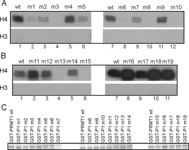
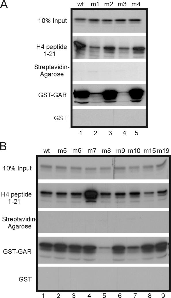
Protein arginine methyltransferase 1 (PRMT1) is an arginine-specific protein methyltransferase that methylates a number of proteins involved in transcription and other aspects of RNA metabolism. Its role as a transcriptional coactivator for nuclear receptors involves its ability to bind to other coactivators, such as glucocorticoid receptor-interacting protein 1 (GRIP1), as well as its ability to methylate histone H4 and coactivators such as peroxisome proliferator-activated receptor gamma coactivator-1alpha. Its ability to form homodimers or higher-order homo-oligomers also is important for its methyltransferase activity. To understand the function of PRMT1 further, 19 surface residues were mutated, based on the crystal structure of PRMT1. Mutants were characterized for their ability to bind and methylate various substrates, form homodimers, bind GRIP1, and function as a coactivator for the androgen receptor in cooperation with GRIP1. We identified specific surface residues that are important for methylation substrate specificity and binding of substrates, for dimerization/oligomerization, and for coactivator function. This analysis also revealed functional relationships between the various activities of PRMT1. Mutants that did not dimerize well had poor methyltransferase activity and coactivator function. However, surprisingly, all dimerization mutants exhibited increased GRIP1 binding, suggesting that the essential PRMT1 coactivator function of binding to GRIP1 may require dissociation of PRMT1 dimers or oligomers. Three different mutants with altered substrate specificity had widely varying coactivator activity levels, suggesting that methylation of specific substrates is important for coactivator function. Finally, identification of several mutants that exhibited reduced coactivator function but appeared normal in all other activities tested, and finding one mutant with very little methyltransferase activity but normal coactivator function, suggested that these mutated surface residues may be involved in currently unknown protein-protein interactions that are important for coactivator function.


Figures

Fig. 1
Fig. 1
Locations of surface mutations on PRMT1. Upper panels show crystal structure of PRMT1 monomer viewed from opposite sides of the protein. Lower panel shows the structure of a homodimer with arrows indicating the dimer interface. Acidic regions are shown in red, while basic regions are blue. Locations of the dimerization arm, docking site for the dimerization arm, active site (where AdoMet and the substrate Arg residue bind) are also indicated. Open arrowheads indicate locations of some surface grooves and bound GAR peptides (curving green cylinders) were observed binding in some of the grooves. Sequence of the GAR peptide (called R3) is shown (15). Acidic PRMT1 surface residues that were mutated to Lys or Arg in this study are designated with abbreviations such as M1 etc, and their locations are indicated with thin black arrows. For each mutant the corresponding amino acid substitutions are given (list). Adapted from Zhang and Cheng (6).
Fig. 2
Fig. 2
Methyltransferase activity of PRMT1 mutants with histone H4. (A & B) Histone H4 or H3 (5 μg) was incubated with approximately 1 μg of each GST-PRMT1 mutant and [3H]AdoMet. Methylated products were observed by SDS-PAGE and autoflurography. (C) Recombinant GST-PRMT1 mutant proteins were observed by SDS-PAGE and staining with Coomassie Blue. Results shown are representative of at least three independent experiments.
Fig. 3
Fig. 3
Methyltransferase activity of PRMT1 mutants with hypomethylated cell extracts. Hypomethylated mouse embryonic fibroblast extracts were incubated with 1 μg of each GST-PRMT1 mutant and [3H]AdoMet. Methylated proteins were separated by SDS-PAGE and visualized by fluorography. Positions of molecular weight markers are indicated on the left. Results shown are representative of at least two independent experiments.
Fig. 4
Fig. 4
Differential methyltransferase activity of PRMT1 mutants with histone H4 vs. GST-GAR. Histone H4, histone H3, GST-GAR or GST (5 μg) was incubated with approximately 1 μg of each GST-PRMT1 mutant and [3H]AdoMet. Reaction products were resolved by SDS-PAGE and visualized by autoflurography. Results shown are representative of three independent experiments.
Fig. 5
Fig. 5
Binding of PRMT1 mutants to histone H4 and GST-GAR. In vitro synthesized, 35S-labeled PRMT1 mutants were incubated with streptavidin-agarose beads containing bound biotin-tagged histone H4 peptide, streptavidin agarose beads alone, or GST-GAR or GST bound to glutathione-agarose beads. Bound PRMT1 was analyzed by SDS-PAGE and autoradiography. Results shown are representative of three independent experiments.
Fig. 6
Fig. 6
Homo-dimerization and binding to GST-GRIP1-AD2 by PRMT1 mutants. GST pull-down assays were performed with GST, GST-PRMT1 (wild type) or GST-GRIP1-AD2 (GRIP1 amino acids 1122−1462) and in vitro synthesized 35S-labeled PRMT1 (wild type or mutants). Bound proteins were analyzed by SDS-PAGE and autoradiography. Results shown are representative of four independent experiments.
Fig. 7
Fig. 7
Coactivator activity of PRMT1 with GRIP1 and androgen receptor. (A) CV1 cells were transiently transfected with MMTV-LUC reporter plasmid (125 ng) and expression vectors encoding AR (10 ng) , GRIP1 (50 ng) and PRMT1 (200 ng). Transfected cells were grown in culture medium with 20 nM DHT and extracts of harvested cells were tested for luciferase activity. Results shown are representative of at least three independent experiments. (B) Expression of PRMT1 mutants was determined in COS7 cells after transfection with 2.5 μg PRMT1 mutant expression vectors. PRMT1 was detected by immunoblot with anti-HA antibody.
Fig. 8
Fig. 8
PRMT1 mutations grouped by phenotype. Opposite side views of a ribbon diagram of a PRMT1 dimer are shown. The location of each class of mutation on the PRMT1 surface is indicated by a different color, as indicated at the bottom of the figure. Circles of corresponding colors are used to show localization of mutations with similar phenotypes within a specific region of the PRMT1 surface.

References

    1. Bedford MT, Richard S. Arginine methylation an emerging regulator of protein function. Mol Cell. 2005;18:263–272. - PubMed
    1. Lee DY, Teyssier C, Strahl BD, Stallcup MR. Role of protein methylation in regulation of transcription. Endocr Rev. 2005;26:147–170. - PubMed
    1. McBride AE, Silver PA. State of the arg: protein methylation at arginine comes of age. Cell. 2001;106:5–8. - PubMed
    1. Lee J, Sayegh J, Daniel J, Clarke S, Bedford MT. PRMT8, a new membrane-bound tissue-specific member of the protein arginine methyltransferase family. J Biol Chem. 2005;280:32890–32896. - PubMed
    1. Zhang X, Zhou L, Cheng X. Crystal structure of the conserved core of protein arginine methyltransferase PRMT3. EMBO J. 2000;19:3509–3519. - PMC - PubMed

Publication types

MeSH terms