. 2007 Nov;56(11):1733-42.
doi: 10.1007/s00262-007-0317-0. Epub 2007 Apr 11.

Human NK cell infusions prolong survival of metastatic human neuroblastoma-bearing NOD/scid mice

Affiliations

Human NK cell infusions prolong survival of metastatic human neuroblastoma-bearing NOD/scid mice

Roberta Castriconi et al. Cancer Immunol Immunother. 2007 Nov.

Abstract

Aim: Several lines of evidence suggest that NK cell immunotherapy may represent a successful approach in neuroblastoma (NB) patients refractory to conventional therapy. However, homing properties, safety and therapeutic efficacy of NK cell infusions need to be evaluated in a suitable preclinical murine NB model.

Materials and methods: Here, the therapeutic efficacy of NK cell infusions in the presence or absence of NK-activating cytokines have been evaluated in a NB metastatic model set up in NOD/scid mice, that display reduced functional activity of endogenous NK cells.

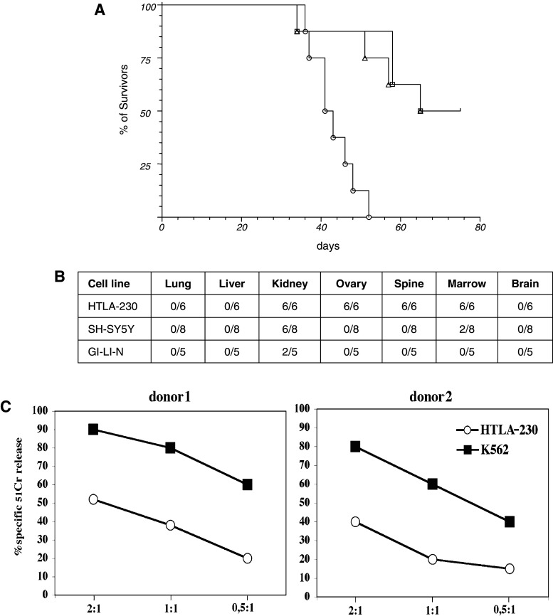
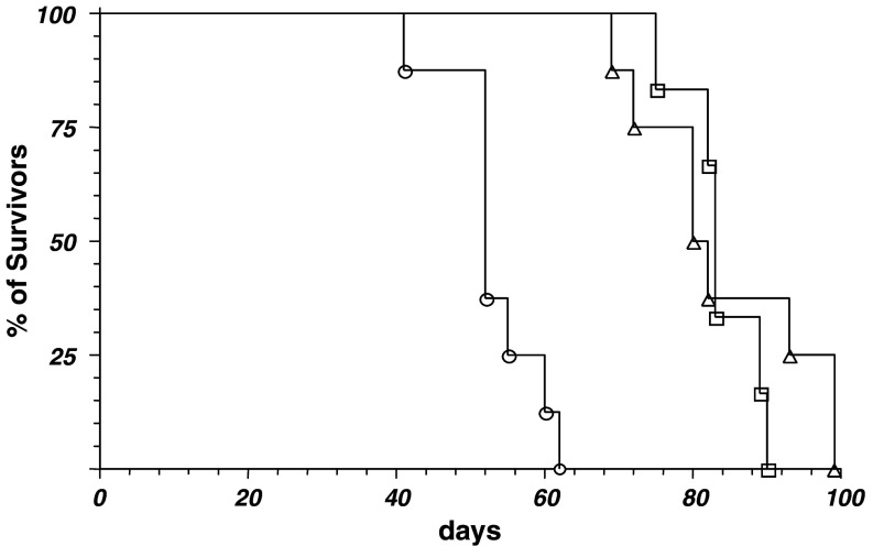
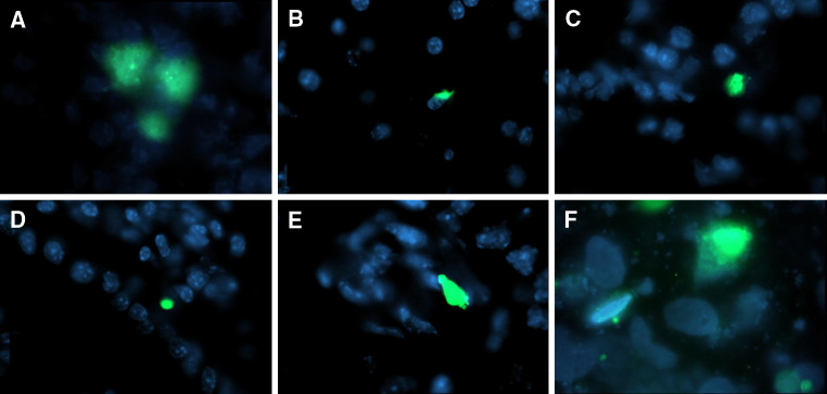
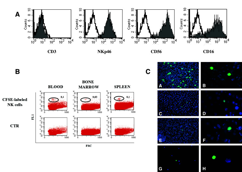
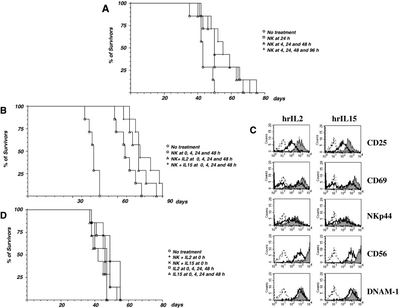
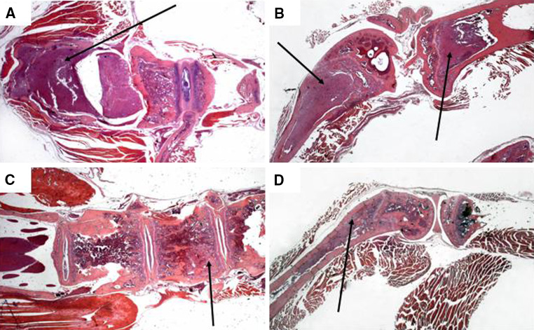
Results: In NOD/scid mice the injected NB cells rapidly reached all the typical sites of metastatization, including bone marrow. Infusion of polyclonal IL2-activated NK cells was followed by dissemination of these cells into various tissues including those colonized by metastatic NB cells. The early repeated injection of IL2-activated NK cells in NB-bearing NOD/scid mice significantly increased the mean survival time, which was associated with a reduced bone marrow infiltration. The therapeutic effect was further enhanced by low doses of human recombinant IL2 or IL15.

Conclusion: Our results indicate that NK-based adoptive immunotherapy can represent a valuable adjuvant in the treatment of properly selected NB patients presenting with metastatic disease, if performed in a minimal residual disease setting.


Figures

Fig. 1
Fig. 1
Tumorigenicity of NB cell lines in NOD/scid mice. a HTLA-230 (circle), SH-SY-5Y (square) and GI-LI-N (triangle) cell lines were iv injected in 5-week-old NOD/scid mice (8 mice/group) at the dose of 3 × 106 in 100 μL serum free medium. Mice were monitored and terminated when bulky disease was apparent (varying from 30 to 60 days) or at day 75 post injection. Terminated animals were next evaluated for the presence of metastasis in the various organs. b Presence of metastasis in the different organs evaluated when the mice showed in a were sacrificed. The experiment was performed twice with similar results. c Susceptibility of HTLA-230 cell line to NK-mediated cytolic activity. Effector cells represented by activated NK cell populations derived from two representative healthy donors were tested against the indicated cell lines at different effector to target (E:T) ratios. The results are representative of three independent experiments; the standard deviation of the mean of the triplicates was <5%. All the therapeutic experiments were performed with polyclonal NK cells derived from donor #1
Fig. 2
Fig. 2
Age-dependent tumorigenicity of HTLA-230 cells. 1 × 10HTLA-230 cells were iv injected in groups (6 mice/group) of 4 (circle), 8 (triangle) and 12 (square) week-old NOD/scid mice. Animals were monitored and terminated at the appearance of bulky disease. The experiment was performed twice with similar results
Fig. 3
Fig. 3
Fate of injected HTLA-230 cell line; 4 × 10CFSE-labeled HTLA-230 cells were iv injected in groups of 3 NOD/scid mice. Animals were terminated at different times pi, autopsy performed, organs removed and snap frozen in liquid nitrogen. Cryostat sections (6 μm thick) were then analyzed by fluorescence microscopy. Sections of organs removed 4 h pi are shown; CFSE-labeled HTLA-230 (green), organ cells (blue). a Lung, b liver, c kidney, d gut, e ovary, f bone marrow
Fig. 4
Fig. 4
Fate of injected IL-2 activated NK cells; 4 × 106 CFSE-labeled NK cells were iv injected in groups of 3 NOD/scid mice. Animals were terminated at different times pi. A Cytofluorimetric analysis of in vitro IL-2 activated NK cells demonstrating their purity. B Blood, bone marrow flushed from femurs, and spleen cell suspensions analyzed by flow cytometry. Percentages of CFSE-labeled NK cells (green) with respect to total white blood cell count are indicated. CTR was represented by cells derived from mice injected with unlabelled polyclonal NK cells. C Fluorescence microscopic analysis of cryostat sections of lung (a, b), liver (c, d), kidney (e, f) and bone marrow smears (g, h) at ×20 (a, c, e, g) and ×100 (b, d, f, h) magnification, respectively. Samples were from mice terminated at 24 h pi; CFSE-labeled NK cells (green), organ cells (blue)
Fig. 5
Fig. 5
Therapeutic effects of NK cell infusions. Panel a four groups of 7, 5-week-old, NOD/scid mice were iv injected with 1 × 106 HTLA-230 cells, then animals were treated with medium alone (circle) or with 4 × 10polyclonal NK cells at 24 h pi (square), at 4, 24 and 48 h pi (triangle) or at 4, 24, 48 and 96 h pi (plus sign). Panel b four groups of 7, 5 week-old, NOD/scid mice were iv injected with 1 × 106 HTLA-230 cells, then animals were treated at 0, 4, 24 and 48 h pi with medium alone (circle), with 4 × 106 polyclonal NK cells (square), with 4 × 106 polyclonal NK cells + hrIL2 (triangle) or 4 × 106 polyclonal NK cells + hrIL15 (plus sign). Panel c IL2-activated NK cells from donor #1 were cultured in the absence or in the presence of rhIL2 (100 IU/mL) and rhIL15 (50 ng/mL). After 3 days, cells were stained with mAbs specific for the indicated molecules, followed by PE-conjugated goat anti-mouse isotype-specific second reagent and analyzed by flow cytometry. Sketched profiles cells incubated with second reagent alone, white profiles cells cultured without cytokines, grey profiles cells cultured with the indicated cytokine. Panel d Five groups of 7, 5-week-old, NOD/scid mice were iv injected with 1 × 106 HTLA-230 cells, then animals were treated with medium alone (circle), with 4 × 106 polyclonal NK cells + hr IL2 at 0 h pi (square), with 4 × 106 polyclonal NK cells + hr IL15 at 0 hr pi (triangle), with 100 ng/mouse hrIL2 at 0, 4, 24 and 48 h pi (plus sign) or with 50 ng/mouse hrIL15 at 0, 4, 24 and 48 h pi (multiple sign). All the experiments were performed twice with similar results
Fig. 6
Fig. 6
Histological analysis of untreated and NK + cytokine-treated mice. Hematoxylin-eosin stained sections of spinal cord and vertebrae (left) and femur and tibia (right) from untreated (a, b) and NK cell + cytokine-treated (c, d) tumor-bearing NOD/scid mice. Magnification is ×12.5. Panel a arrow indicates neuroblastoma cells grown outside the vertebral bone towards the spinal cord. Panel b arrows indicate neuroblastoma cells grown in the femur and tibia bones. In the femur NB infiltration destroyed the bone structure while in the tibia NB growth is confined to the bone marrow. Panel c arrow indicates neuroblastoma cells inside the vertebral bone, on the left side the spinal cord appears completely free from metastatic invasion. Panel d arrow indicates neuroblastoma cells grown inside the femur bone marrow without destruction of the bone structure, the tibia bone marrow appears free of neuroblastoma cells

References

    1. Brodeur GM. Neuroblastoma: biological insights into a clinical enigma. Nat Rev Cancer. 2003;3:203–216. doi: 10.1038/nrc1014. - DOI - PubMed
    1. Schwab M, Westermann F, Hero B, Berthold F. Neuroblastoma: biology and molecular and chromosomal pathology. Lancet Oncol. 2003;4:472–480. doi: 10.1016/S1470-2045(03)01166-5. - DOI - PubMed
    1. Brodeur GM, Maris JM. Neuroblastoma. In: Pizzo PA, Poplack DG, editors. Principles and practice of pediatric oncology. Philadelphia: Lippincott–Raven; 2001. pp. 895–937.
    1. Carlsen NL, Castel V, Castelberry RP, De Bernardi B, Evans AE, Favrot M, Hedborg F. Revisions of the international criteria for neuroblastoma diagnosis, staging, and response to treatment. J Clin Oncol. 1993;11:1466–1477. - PubMed
    1. Maris JM, Matthay KK. Molecular biology of neuroblastoma. J Clin Oncol. 1999;17:2264–2279. - PubMed

Publication types

Substances