Review
. 2007:30:339-65.
doi: 10.1146/annurev.neuro.29.051605.112917.

The micromachinery of mechanotransduction in hair cells

Affiliations
Review

The micromachinery of mechanotransduction in hair cells

Melissa A Vollrath et al. Annu Rev Neurosci. 2007.

Abstract

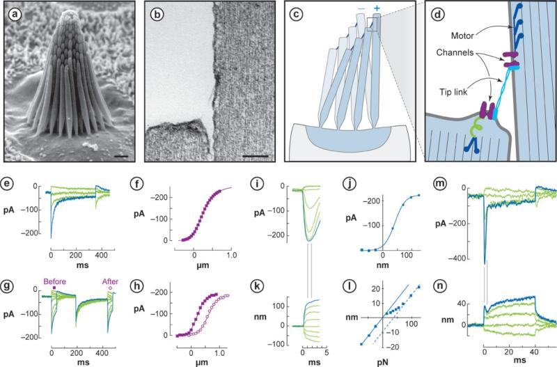
Mechanical stimuli generated by head movements and changes in sound pressure are detected by hair cells with amazing speed and sensitivity. The mechanosensitive organelle, the hair bundle, is a highly elaborated structure of actin-based stereocilia arranged in precise rows of increasing height. Extracellular linkages contribute to its cohesion and convey forces to mechanically gated channels. Channel opening is nearly instantaneous and is followed by a process of sensory adaptation that keeps the channels poised in their most sensitive range. This process is served by motors, scaffolds, and homeostatic mechanisms. The molecular constituents of this process are rapidly being elucidated, especially by the discovery of deafness genes and antibody targets.


Figures

Figure 1
Figure 1
Hair cell anatomy and physiology. (a) Hair bundle in a bullfrog saccule, comprising ~60 stereocilia and a single kinocilium (adjacent to tallest stereocilia) (scale bar = 1 μm). (b) Two stereocilia and the tip link extending between them (scale bar = 0.1 μm). (c) Deflection of hair bundle. Stereocilia tips remain in contact but shear with deflection. (d) Schematic model of transduction. Shearing with positive deflection increases tension in tip links, which pull open a transduction channel at each end. Myosin motors slip or climb to restore resting tension. An elastic gating spring likely exists between a channel and the actin cytoskeleton. (e) Mouse utricular hair cell transduction currents. Step deflections (not shown) from –0.3 to +0.7 μm elicit inward currents that adapt over 10–50 ms. (f) Activation or I(X) curve shows an operating range of ~0.7 μm, with ~12% of channels open at rest. Inward current is shown here as upward to reflect open probability. (g) Multiple short stimulus steps (not shown) map the I(X) curve before (square) and after (open circle) a 250-ms, 0.4-μm adapting step. (h) Adaptation occurs as a shift of the activation curve in the stimulus direction to an extent of 80%–90% of the stimulus amplitude. (i) Bullfrog saccular hair cell transduction currents, measured 1 ms after onset of a force stimulus delivered by an optical trap. (j) The I(X) curve is very narrow (~0.1 μm) when measured with fast stimuli. (k) Corresponding bundle movements elicited by the force stimuli. (l) Force-displacement relation measured 1 ms after onset of the stimulus. The bundle shows simple elastic movements for stimuli where channels are all closed or all open but is more compliant over the range of channel opening. (m) Fast and slow adaptation in a bullfrog hair cell. Positive force steps (not shown) elicit rapid channel opening followed by adaptation occurring in two phases, a fast phase of 1–3 ms and a slow phase of 10–50 ms. (n) Corresponding bundle movements reveal correlates of adaptation: a quick negative movement associated with fast adaptation and a slower positive movement with slow adaptation.
Figure 2
Figure 2
Proteins associated with links between stereocilia. (a) Position of different links between stereocilia. (b) Links at tips of stereocilia. (c) Protein domain structures. CA, cadherin domain; tm, transmembrane domain; FN3, fibronectin type 3 domain; PTPc, protein tyrosine phosphatase catalytic domain; LamNT, laminin N-terminal domain; EGF lam, laminin-type epidermal growth factor–like domain; LamG, laminin G domain; calx beta, Ca2+ exchanger/integrin-beta4 domain; EPTP, epitempin-like domain; GPS, G protein–coupled receptor proteolytic site domain; GPCR7tm, G protein–coupled receptor seven-transmembrane domain.
Figure 3
Figure 3
Proteins associated with adaptation. Myosin-1c is located in the hair bundle, especially in the distal third, and is most concentrated at either end of the tip link. Myosin-7a is found throughout the bundle. Both are found in the vesicle-rich pericuticular zone (pz) between the cuticular plate (cp) and the membrane. IQ, regulatory light-chain-binding domain; HDACI, histone deacetylase interacting domain; EFH, EF hand (Ca2+-binding) domain; cc, coiled-coil domain; MyTH4, myosin tail homology domain 4; FERM, 4.1/ezrin/radixin/moesin-like domain; SH3, Src homology 3 domain; PDZ, PSD-95/Dlg/ZO-1-like domain.
Figure 4
Figure 4
Proteins associated with ion homeostasis. All are located in stereocilia; the Ca2+-binding proteins calbindin and oncomodulin are in the soma as well. ATPase, cation transporter ATPase domain; hydrolase, haloacid dehalogenase-like hydrolase domain.
Figure 5
Figure 5
Protein candidates for the mechanosensory transduction channel. The channel is located most likely at either end of the tip link. A, ankyrin domain. Lightly-shaded transmembrane domains are predicted by hydropathy but not common to other members of the TRP channel family.

References

    1. Adato A, Lefevre G, Delprat B, Michel V, Michalski N, et al. Usherin, the defective protein in Usher syndrome type IIA, is likely to be a component of interstereocilia ankle links in the inner ear sensory cells. Hum. Mol. Genet. 2005a;14:3921–32. - PubMed
    1. Adato A, Michel V, Kikkawa Y, Reiners J, Alagramam KN, et al. Interactions in the network of Usher syndrome type 1 proteins. Hum. Mol. Genet. 2005b;14:347–56. - PubMed
    1. Ahmed ZM, Goodyear R, Riazuddin S, Lagziel A, Legan PK, et al. The tip-link antigen, a protein associated with the transduction complex of sensory hair cells, is protocadherin-15. J. Neurosci. 2006;26:7022–34. - PMC - PubMed
    1. Ahmed ZM, Riazuddin S, Bernstein SL, Ahmed Z, Khan S, et al. Mutations of the protocadherin gene PCDH15 cause Usher syndrome type 1F. Am. J. Hum. Genet. 2001;69:25–34. - PMC - PubMed
    1. Alagramam KN, Kwon HY, Cacheiro NL, Stubbs L, Wright CG, et al. A new mouse insertional mutation that causes sensorineural deafness and vestibular defects. Genetics. 1999;152:1691–99. - PMC - PubMed

Publication types

MeSH terms

LinkOut - more resources