. 2007 May 15;313(9):1820-9.
doi: 10.1016/j.yexcr.2007.02.031. Epub 2007 Mar 12.

Synthetic nanostructures inducing differentiation of human mesenchymal stem cells into neuronal lineage

Affiliations

Synthetic nanostructures inducing differentiation of human mesenchymal stem cells into neuronal lineage

Evelyn K F Yim et al. Exp Cell Res. .

Abstract

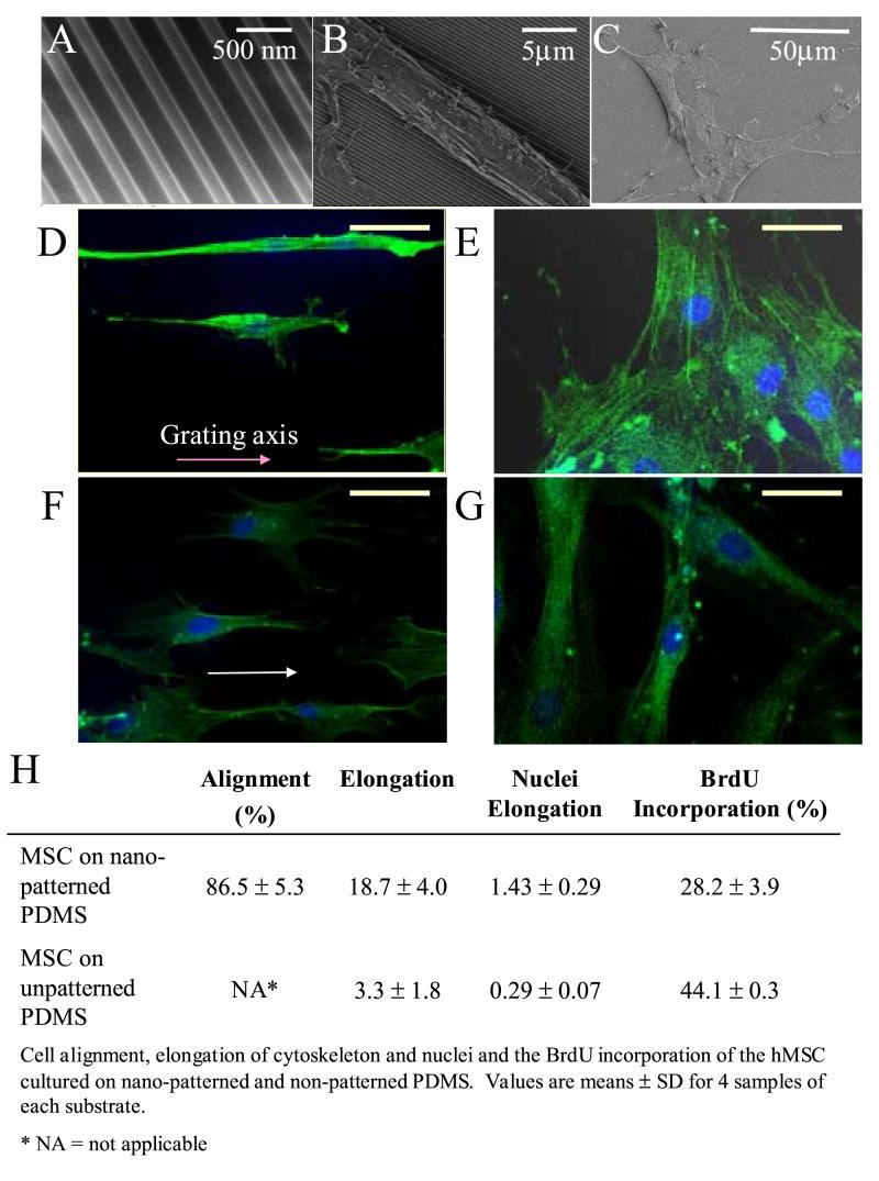
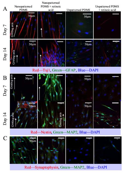
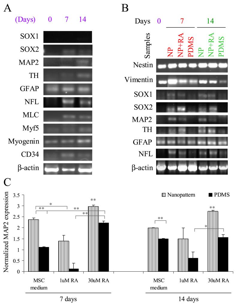
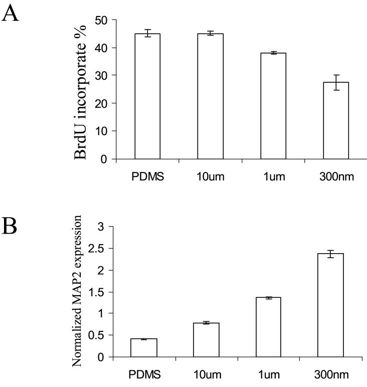
Human mesenchymal stem cells (hMSCs) have been shown to trans-differentiate into neuronal-like cells by culture in neuronal induction media, although the mechanism is not well understood. Topography can also influence cellular responses including enhanced differentiation of progenitor cells. As extracellular matrix (ECM) in vivo comprises topography in the nanoscale, we hypothesize that nanotopography could influence stem cell differentiation into specific non-default pathways, such as transdifferentiation of hMSCs. Differentiation and proliferation of hMSCs were studied on nanogratings of 350 nm width. Cytoskeleton and nuclei of hMSCs were aligned and elongated along the nanogratings. Gene profiling and immunostaining showed significant up-regulation of neuronal markers such as microtubule-associated protein 2 (MAP2) compared to unpatterned and micropatterned controls. The combination of nanotopography and biochemical cues such as retinoic acid further enhanced the up-regulation of neuronal marker expressions, but nanotopography showed a stronger effect compared to retinoic acid alone on unpatterned surface. This study demonstrated the significance of nanotopography in directing differentiation of adult stem cells.


Figures

Figure 1
Figure 1
Changes in morphology and proliferation of human mesenchymal stem cells (hMSCs) cultured on nano-gratings. Scanning electron micrographs of (A) PDMS nano-patterned by replica molding; hMSCs cultured on (B) nano-patterned PDMS and (C) unpatterned PDMS. Confocal micrographs of F-actin-stained hMSCs on (D) nano-patterned PDMS and (E) unpatterned PDPS in hMSC proliferation medium; (F) nano-patterned PDMS and (G) unpatterned PDMS cultured in presence of 1μM of retinoic acid (RA). Bar = 500nm for A, 5 μm for B, 50 μm for C-G. (H) Cell alignment, elongation of cytoskeleton and nuclei and the BrdU incorporation of the hMSCs cultured on nano-patterned and unpatterned PDMS. Values are means ± SD for 4 samples of each substrate. * NA = not applicable
Figure 2
Figure 2
Immunofluorescent staining of (A) Tuj1 and GFAP, (B) MAP2 and nestin and (C) synaptophysin and MAP2 of hMSCs cultured on nano-patterned PDMS, nano-patterned PDMS in the presence of retinoic acid (RA), unpatterned PDMS and unpatterned PDMS in the presence of RA. (A) Tuj1 is shown in red, GFAP in green; (B) nestin is shown in red, MAP2 in green and (C) synaptophysin is shown in red and marked by arrows, while MAP2 is shown in green. In all panels the DAPI nuclei counter-stain is shown in blue, bar = 50 μm. The direction of the gratings on the nano-patterned PDMS is indicated with a white arrow. Images were taken at representative areas of the samples.
Figure 3
Figure 3
(A) Multi-lineage gene expression analysis of hMSCs cultured on nano-patterned PDMS and the unpatterned control in proliferation medium at day 7 and day 14. (B) Neuronal gene markers analysis of hMSCs cultured on nano-patterned (NP) PDMS in proliferation medium or in the presence of retinoic acid (RA), and unpatterned PDMS (PDMS). hMSCs cultured on TCPS were used as the day 0 control. (C) Quantitative analysis of MAP2 expression in hMSCs cultured on nano-patterned PDMS in proliferation medium or in the presence of RA and on unpatterned PDMS and tissue culture polystyrene (TCPS) surface control on day 7 and day 14. * p < 0.01 and ** p < 0.001, two-tailed t-test. The expression of MAP2 was first normalized with the expression of endogenous control β-actin, then normalized with the MAP2 expression of unpatterned hMSC control at day 7.
Figure 4
Figure 4
Proliferation and differentiation of the hMSCs on gratings with various widths. (A) Brdu incorporation percentage after a 4-hour pre-fix incubation and (B) quantitative analysis of MAP2 expression in hMSCs cultured on gratings with widths of 350nm, 1μm and 10μm.

References

    1. Dalby MJ, McCloy D, Robertson M, Agheli H, Sutherland D, Affrossman S, Oreffo RO. Osteoprogenitor response to semi-ordered and random nanotopographies. Biomaterials. 2006;27:2980–2987. - PubMed
    1. Silva GA, Czeisler C, Niece KL, Beniash E, Harrington DA, Kessler JA, Stupp SI. Selective differentiation of neural progenitor cells by high-epitope density nanofibers. Science. 2004;303:1352–1355. - PubMed
    1. Curtis A, Wilkinson C. Nantotechniques and approaches in biotechnology. Trends Biotechnol. 2001;19:97–101. - PubMed
    1. Curtis AS, Wilkinson CD. Reactions of cells to topography. J Biomater Sci Polym Ed. 1998;9:1313–1329. - PubMed
    1. Wilkinson CDW. Nanostructures in Biology. Microelectron. Eng. 1995;27:61–65.

Publication types

MeSH terms