. 2007 Jun 1;282(22):16054-61.
doi: 10.1074/jbc.M611392200. Epub 2007 Apr 11.

RNA binding-independent dimerization of adenosine deaminases acting on RNA and dominant negative effects of nonfunctional subunits on dimer functions

Affiliations

RNA binding-independent dimerization of adenosine deaminases acting on RNA and dominant negative effects of nonfunctional subunits on dimer functions

Louis Valente et al. J Biol Chem. .

Abstract

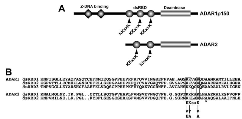
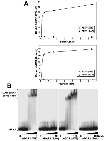
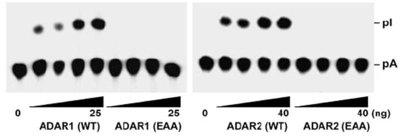
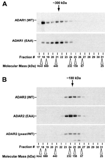
RNA editing that converts adenosine to inosine in double-stranded RNA (dsRNA) is mediated by adenosine deaminases acting on RNA (ADAR). ADAR1 and ADAR2 form respective homodimers, and this association is essential for their enzymatic activities. In this investigation, we set out experiments aiming to determine whether formation of the homodimer complex is mediated by an amino acid interface made through protein-protein interactions of two monomers or via binding of the two subunits to a dsRNA substrate. Point mutations were created in the dsRNA binding domains (dsRBDs) that abolished all RNA binding, as tested for two classes of ADAR ligands, long and short dsRNA. The mutant ADAR dimer complexes were intact, as demonstrated by their ability to co-purify in a sequential affinity-tagged purification and also by their elution at the dimeric fraction position on a size fractionation column. Our results demonstrated ADAR dimerization independent of their binding to dsRNA, establishing the importance of protein-protein interactions for dimer formation. As expected, these mutant ADARs could no longer perform their catalytic function due to the loss in substrate binding. Surprisingly, a chimeric dimer consisting of one RNA binding mutant monomer and a wild type partner still abolished its ability to bind and edit its substrate, indicating that ADAR dimers require two subunits with functional dsRBDs for binding to a dsRNA substrate and then for editing activity to occur.


Figures

FIGURE 1
FIGURE 1. ADAR double-stranded RNA binding domains and the KKXXK motif
A, domain structure of ADAR1 and ADAR2 indicating Z-DNA binding domains (triangles), dsRBD (circles), and the C-terminal deaminase domain (rectangle). The KKXXK motif in each dsRBD is indicated. B, sequence alignment of the ADAR1 (top) and ADAR2 (bottom) dsRBD displaying the homology around the KKXXK motif and the mutated lysines to EAXXA (termed EAA). The asterisk denotes an alanine mutated in other studies.
FIGURE 2
FIGURE 2. ADAR dsRBD mutation abolishes binding to long and short dsRNA
A, binding of recombinant proteins to c-Myc dsRNA (583 base pairs) was analyzed by a nitrocellulose filter binding assay. 10 ng of purified ADAR1 (WT and EAA) (top panel) and ADAR2 (WT and EAA) (bottom panel) protein were incubated at 30 °C for 5 min in triplicate with various concentrations of dsRNA substrate. B, binding of recombinant ADAR1 (WT and EAA) and ADAR2 (WT and EAA) proteins to short dsRNA was examined by electrophoretic mobility shift assay. Binding of ADAR1 proteins up to 50-fold over Kd values for wild type (10 nm) and ADAR2 proteins, up to 10-fold over Kd values for wild type (100 nm) to 10 pm 32P-labeled 19 base pairs enhanced green fluorescent protein short interfering RNA was analyzed on a native 4.5% polyacrylamide gel.
FIGURE 3
FIGURE 3. dsRBD mutant ADARs completely lack the A → I modification activity
The A → I conversion of c-Myc dsRNA (20 fmol) was monitored with increasing amounts of purified recombinant ADAR1 (WT and EAA) (right panel, 3.13, 6.25, 12.5, and 25 ng) and ADAR2 (WT and EAA) (left panel, 5, 10, 20, and 40 ng) proteins. Following incubation for 1 h at 37 °C, the reaction products were deproteinized, digested with P1 nuclease, and analyzed by thin layer chromatography. pA, 5′-AMP; pI, 5′-IMP.
FIGURE 4
FIGURE 4. Homodimerization of ADAR is independent of dsRNA binding
The ADAR1 (WT and EAA) and ADAR2 (WT and EAA) proteinswere co-expressed in Sf9 cells with its differentially tagged partner. These recombinant proteins were sequentially purified on an anti-FLAGmAb affinity column (left panel) and then on a second TALON affinity column (right panel). Western blotting analysis specific for the first purification using the anti-FLAG M2 mAb indicates the F-tagged protein eluted (left panel) and the anti-His6 mAb reveals the H-tagged protein that is retrieved (right panel), confirming the interaction. Single purified recombinant F-ADAR2 and H-ADAR2 expressed with one tag was included to show the specificity of the two mAbs used forWestern blotting analysis (lanes 1, 2, 7, and 8). These F/H-tagged ADAR1 and ADAR2 oligomeric complexes were alsoidentified with the reciprocal mAb toconfirm the presence of the other monomer subunit partner (not shown). Approximately 10 ng of each purified protein was loaded onto the SDS-polyacrylamide gels.
FIGURE 5
FIGURE 5. Analysis of dsRNA binding mutant ADAR oligomeric complexes by size exclusion chromatography
Recombinant purified ADAR1 (WT and EAA) (A) and ADAR2 (WT and EAA) (B) proteins as well as a yeast-derived wild type ADAR2-purified protein (B, lower panel) were fractionated by Superose 12 gel filtration column chromatography and analyzed by Western blotting using specific mAb for ADAR1 (A) or ADAR2 (B). The positions of molecular 32 size marker protein used as the calibration standards are indicated by open arrowheads. The estimated dimer size for fractions of ADAR1 (~300 kDa) and ADAR2 (~180 kDa) are indicated by black arrows. Running the samples on a denaturing SDS-polyacrylamide gel allowed confirmation of the monomeric size for ADAR1 (150 kDa) and ADAR2 (90 kDa).
FIGURE 6
FIGURE 6. Dominant negative effects of one dsRBD mutant monomer paired with a wild type partner
A, the A → I base modification assay for F/H-ADAR2 (WT/WT), F/H-ADAR2 (EAA/WT), and F/H-ADAR2 (E396A/WT) was carried out with 10 ng of protein for 30 min at 37 °C with 20 fmol of c-Myc dsRNA. B, dsRNA binding assays similar to Fig. 2 were performed with 10 ng of F/H-ADAR2 (WT/WT), F/H-ADAR2 (EAA/WT), F/H-ADAR2 (E396A/WT), and 6.4 nm c-Myc dsRNA. The experiments were normalized to F/H-ADAR2 (WT/WT), and all experiments were done in triplicate.

Similar articles

Cited by

References

    1. Bass BL. Annu Rev Biochem. 2002;71:817–846. - PMC - PubMed
    1. DeCerbo J, Carmichael GG. Curr Opin Cell Biol. 2005;17:302–308. - PubMed
    1. Reenan RA. Trends Genet. 2001;17:53–56. - PubMed
    1. Toth AM, Zhang P, Das S, George CX, Samuel CE. Prog Nucleic Acid Res Mol Biol. 2006;81:369–434. - PubMed
    1. Higuchi M, Single FN, Kohler M, Sommer B, Sprengel R, Seeburg PH. Cell. 1993;75:1361–1370. - PubMed

Publication types