. 2007 May;134(10):1861-71.
doi: 10.1242/dev.002972. Epub 2007 Apr 11.

Dynamic decapentaplegic signaling regulates patterning and adhesion in the Drosophila pupal retina

Affiliations

Dynamic decapentaplegic signaling regulates patterning and adhesion in the Drosophila pupal retina

Julia B Cordero et al. Development. 2007 May.

Abstract

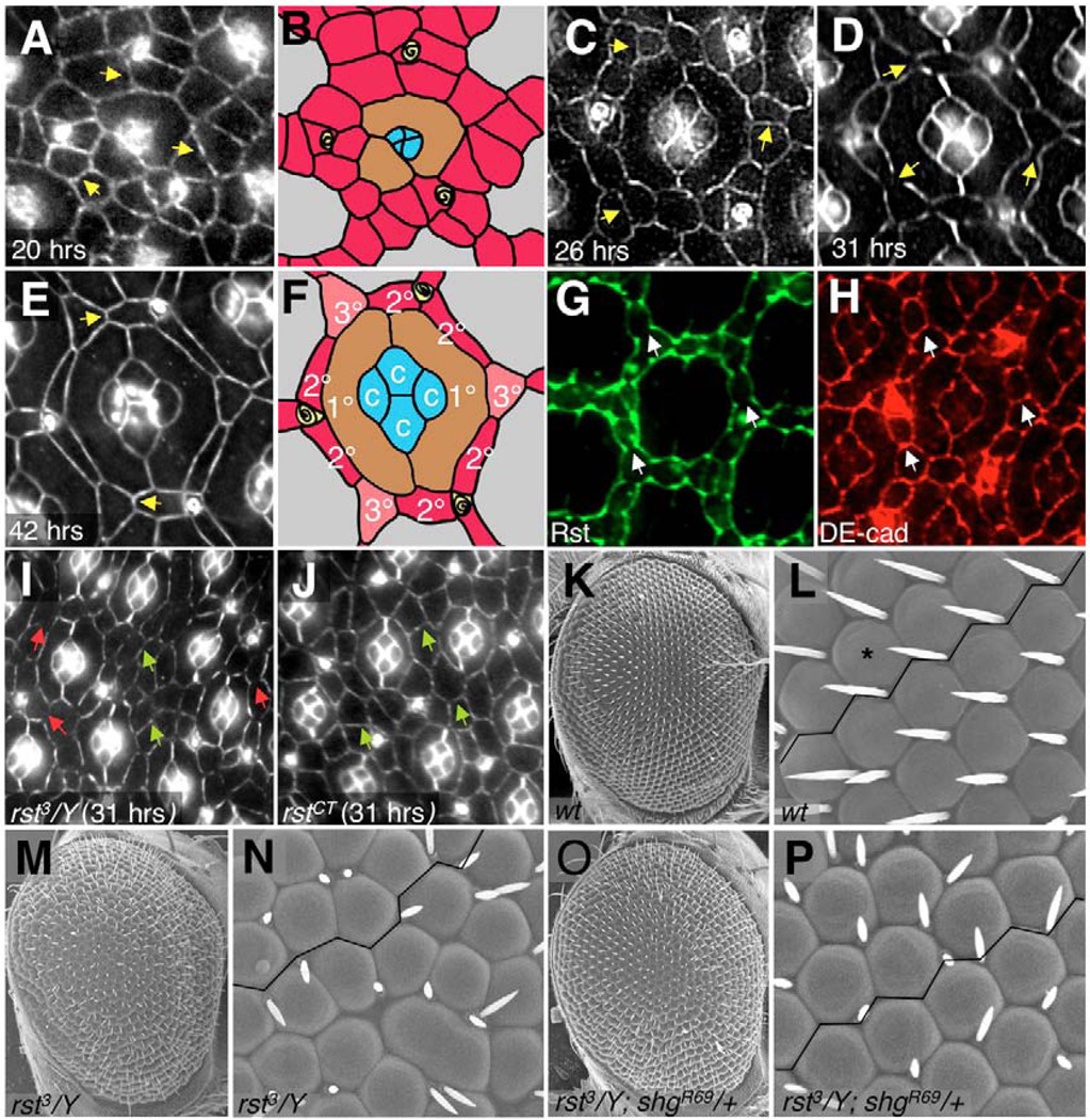
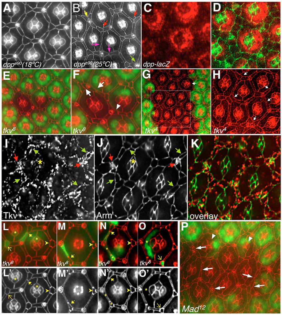
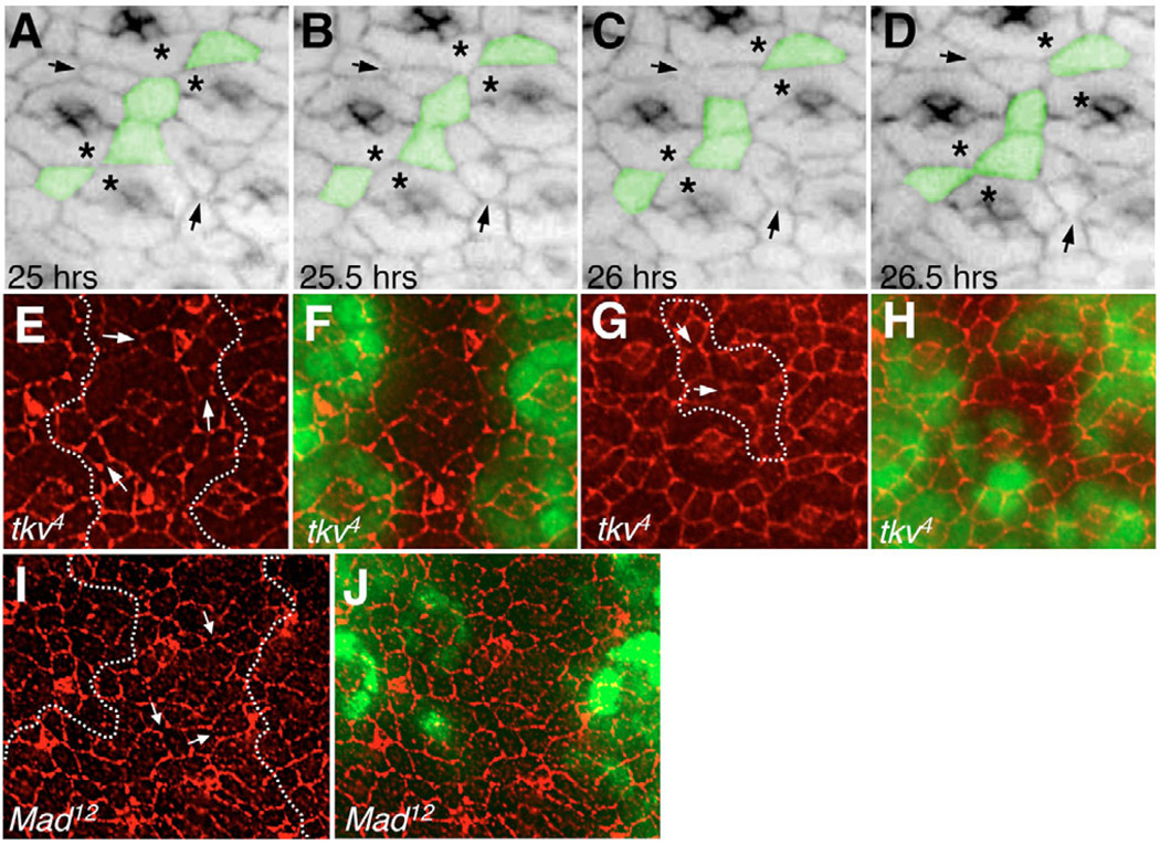
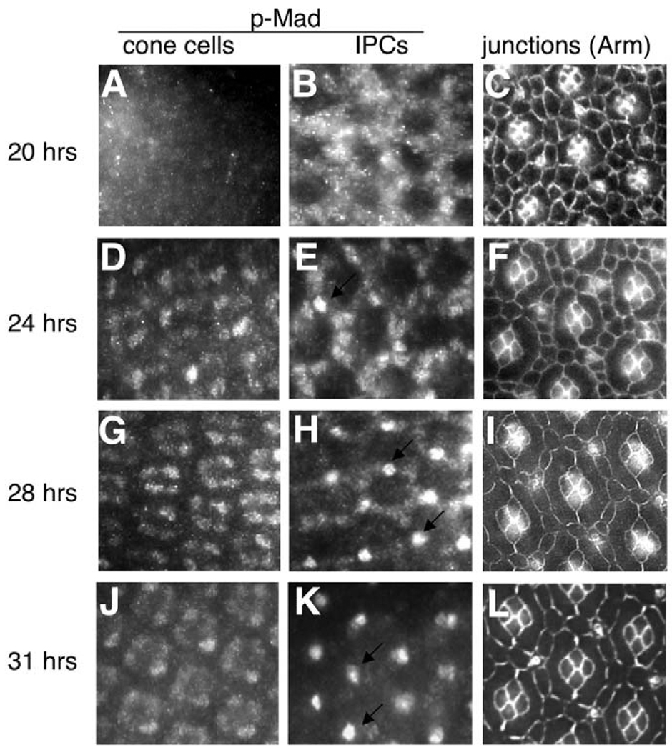
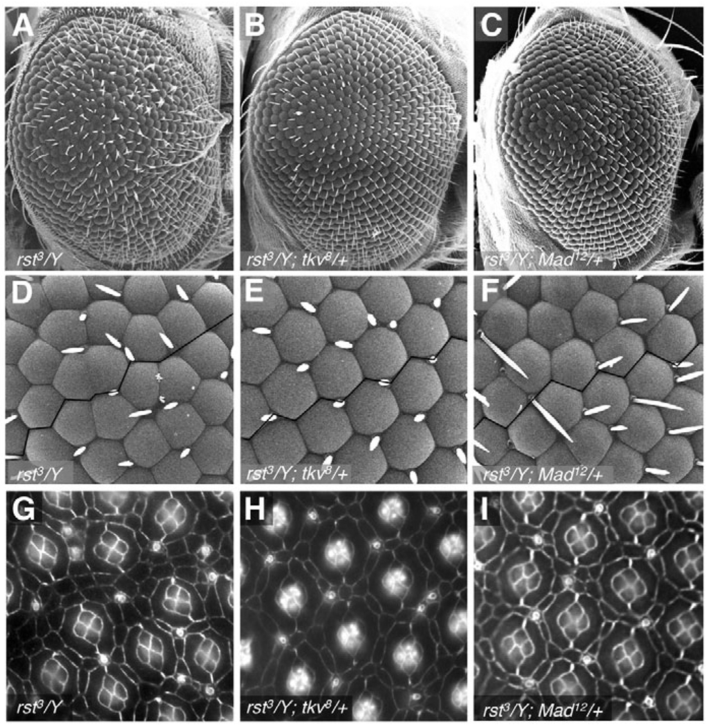
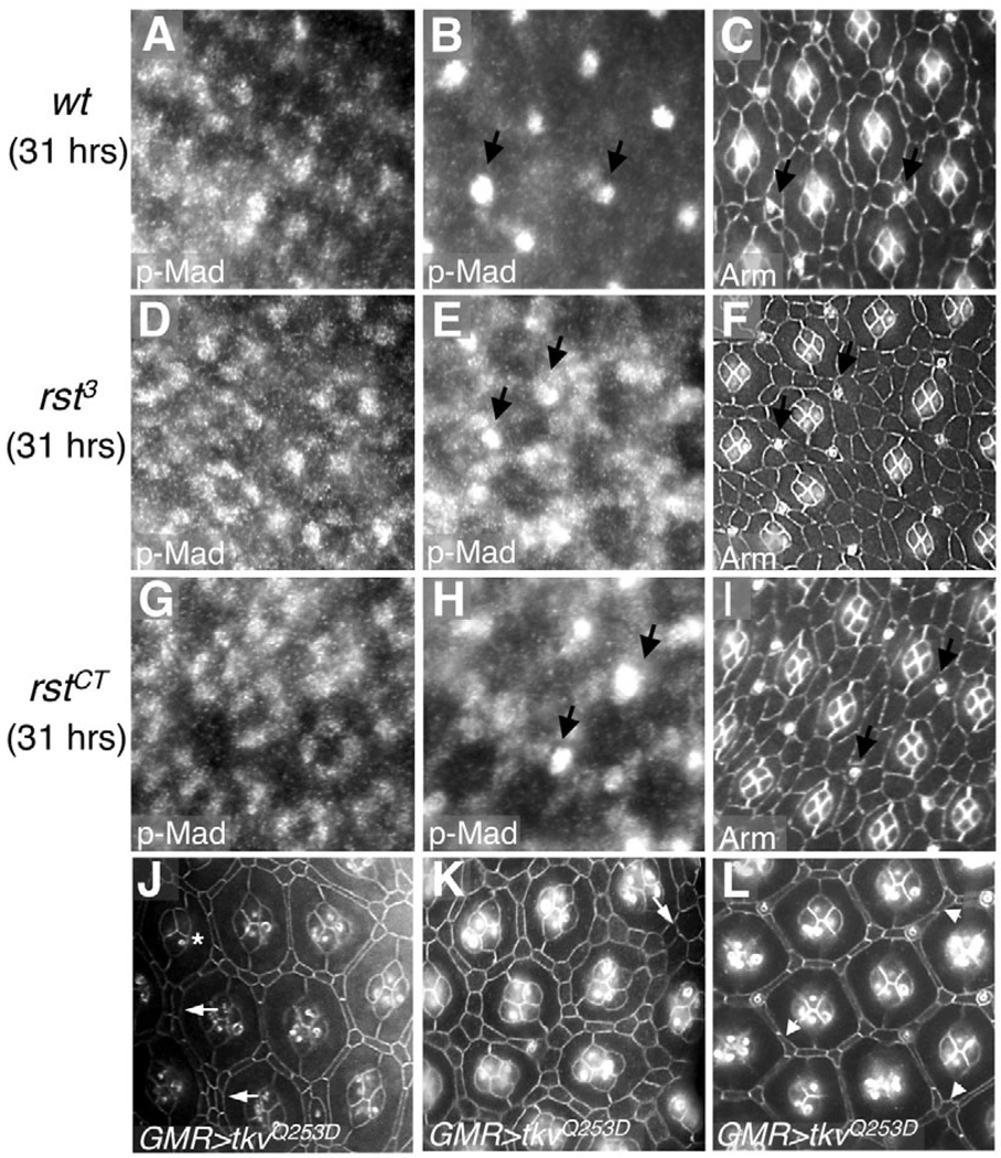
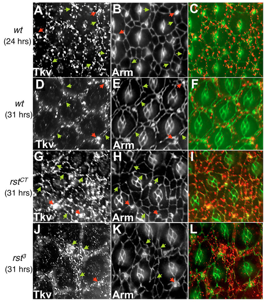
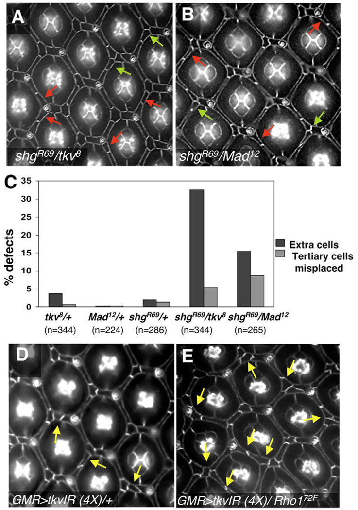
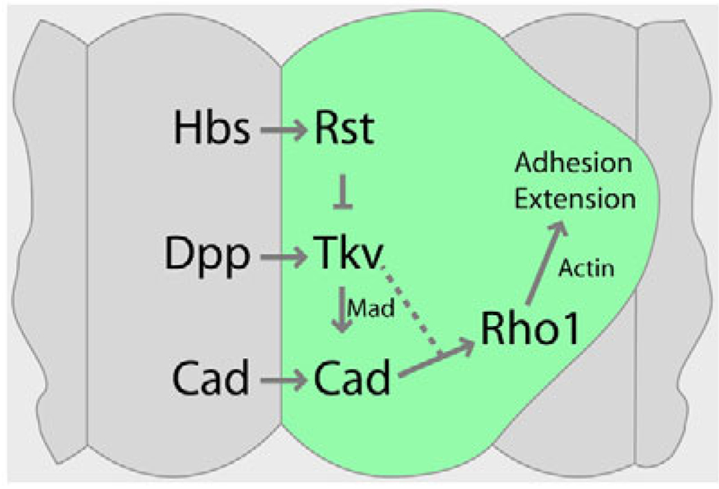
The correct organization of cells within an epithelium is essential for proper tissue and organ morphogenesis. The role of Decapentaplegic/Bone morphogenetic protein (Dpp/BMP) signaling in cellular morphogenesis during epithelial development is poorly understood. In this paper, we used the developing Drosophila pupal retina--looking specifically at the reorganization of glial-like support cells that lie between the retinal ommatidia--to better understand the role of Dpp signaling during epithelial patterning. Our results indicate that Dpp pathway activity is tightly regulated across time in the pupal retina and that epithelial cells in this tissue require Dpp signaling to achieve their correct shape and position within the ommatidial hexagon. These results point to the Dpp pathway as a third component and functional link between two adhesion systems, Hibris-Roughest and DE-cadherin. A balanced interplay between these three systems is essential for epithelial patterning during morphogenesis of the pupal retina. Importantly, we identify a similar functional connection between Dpp activity and DE-cadherin and Rho1 during cell fate determination in the wing, suggesting a broader link between Dpp function and junctional integrity during epithelial development.


Figures

Fig. 1
Fig. 1. Morphogenesis of the Drosophila pupal retina and the role of cell adhesion
(A–F) Time course of retinal development. Apical cell profiles were visualized with anti-Armadillo to highlight adherens junctions. Anterior is to the right; times refers to hours after puparium formation (APF). Unpatterned arrays of IPCs (A,B) sort to single file (C); IPC number continues to decrease as patterning tightens (D) until the final pattern is achieved (E,F). B and F are schematics of the central ommatidium from A and E, respectively; cone cells (c, blue shading), primary pigment cells (1°, brown), IPCs (red), 2° (red), 3° (pink) and bristles (yellow) are indicated. Arrows in A,C,D,E point to IPC:IPC adherens junctions. (G,H) 26 hours APF retina stained with anti-Rst (G) and anti-DE-cadherin (H). Arrows point to the IPC:IPC junctions, where DE-cadherin is expressed and Rst is absent. (I,J) 31 hours APF rst mutant retinas; magnification is reduced to show additional ommatidia. Green arrows point to IPC:IPC junctions that failed to clear (compare with wild type, D). This defect correlated with the failure in mutant IPCs to sort out into single-cell rows, as observed in mutants for the hypomorphic allele rst3, which is subject to position effect variegation (I). Red arrows in I point to rst3 regions where IPCs have sorted out into single layers and have also cleared out their junctions; these are likely to contain normal levels of Rst protein. (K–P) Scanning electron micrographs of adult eyes (genotypes as listed) taken at 180× (K,M,O) and 800× (L,N,P). K and L show a wild-type adult eye; a single ommatidium is indicated with an asterisk. Note the straight ommatidial rows, highlighted by a line drawn between ommatidia. The aberrant ommatidial packing observed in an rst3 eye (M,N) is rescued by removing a single functional copy of shg (O,P).
Fig. 2
Fig. 2. Dpp signaling regulates IPC patterning in the Drosophila pupal retina
All retinas are 42 hours APF and apical membrane profiles are highlighted with anti-Armadillo, except where noted. (A,B) Retinas from animals carrying the temperature-sensitive allele dppe90 raised at the permissive (A) or non-permissive (B) temperature. (B) Mutant retinas show: abnormal IPC:IPC contacts (pink arrows); 3°s, which failed to establish a correct position as a vertex of the hexagon (red arrows); 2°/3°s abnormally arranged around sensory bristles (yellow arrow); and aberrant bristle-bristle contacts (green arrow). (C,D) dpp was expressed in primary pigment cells at 26 hours APF (red). (E,F) tkv8 clone marked by the absence of nuclear GFP (green). (F) Magnification of the clonal tissue in E. Arrows in F point to typical 2°/3°s patterning defects; arrowhead points to ectopic 2°/3°. (G,H) tkv4 clone (cells marked as in E,F). H is a magnified view of the boxed region in G. Arrows in H indicate examples of typical 2°/3° defects. Arrow in G indicates a rare cone cell defect (five versus four). (I–K) Tkv localization at 26 hours APF. (I) Tkv protein was found primarily at the surface of IPCs (green arrows), sensory bristles (red arrows) and at lower levels in cone cells (asterisk). (L–O′) tkv8 single-cell clones were marked by the presence of GFP (green). Full arrows point to cases where tkv mutant 2°s failed to fully expand into their proper niche, as evidenced by their shortened apical profile, while wild-type neighboring 3°s elongated to compensate. Arrowheads point to examples of the apical profile characteristic of wild-type 3°s. Asterisks in N and N′ indicate how neighboring mutant cells typically show normal apical profiles. Thin arrows and asterisks in L,L′,O,O′ point to 3°s, cone cells and primary pigment cells, whose shape was not affected by the absence of Tkv activity. (P) A Mad12 clone marked by the absence of nuclear GFP (green). The arrows point to a subset of the 2°/3°s patterning defects and aberrant bristle-bristle contacts within the clone. Arrowheads indicate rare, abnormal cone cell clusters.
Fig. 3
Fig. 3. Aberrant IPC morphogenesis and unstable IPC-IPC junctions in retinas with reduced Dpp signaling
(A–D) In vivo imaging of a Drosophila retina with reduced Tkv activity [GMR-gal4/+; UAS-αCatenin-GFP/tkv8; UAS-tkv-IR1(2x)/UAS-tkv-IR1(2X)] (see also Movie 2 in the supplementary material). Hours APF are indicated. Pseudo colored in green are examples of IPCs that transiently lose their apical contact and leave primary pigment cells from adjacent ommatidia (asterisks) in direct, aberrant contact. Arrows point to examples of adherens junctions, which disappear as the IPC-IPC surface contact decreases. (E–J) Clones of tkv4 (E–H) and Mad12 (I,J) dissected at 25 hours APF and stained with anti-DE-cadherin (red; E–J). Clonal tissue is marked by the absence of GFP (green; F,H,J) or outlined by dotted lines (E,G,I). Arrows point to IPC-IPC junctions with abnormally low-to-undetectable DE-cadherin staining.
Fig. 4
Fig. 4. Dynamic Dpp signaling activity in the Drosophila pupal retina
(A–L) Dpp pathway activity is visualized in pupal retinas at different developmental stages using anti-p-Mad antibody. (A,D,G,J) p-Mad staining at the level of the cone cell nuclei; (B,E,H,K) p-Mad staining at the level of the IPC nuclei. (C,F,I,L) The maturing IPC pattern from age-matched retinas as development proceeded; cell membranes were stained with anti-Armadillo antibody. Time refers to hours APF. Arrows in E,H,K point to nuclear p-Mad in the sensory bristles.
Fig. 5
Fig. 5. Dpp signaling and Rst work in opposition in the Drosophila pupal retina
Genetic interactions between dpp pathway components and rst. (A–F) Scanning electron micrographs of adult eyes taken at (A–C) 180× and (D–F) 800×. (G–I) 42 hours APF retinas stained with anti-Armadillo antibody. Removing a single functional genomic copy of tkv (B,E,H) or Mad (C,F,I) suppressed the rough eye phenotype of rst3 mutants (A,D,G).
Fig. 6
Fig. 6. Rst function is required to achieve proper Dpp signaling activity in the Drosophila pupal retina
(A,B,D,E,G,H) p-Mad staining in 31 hours APF retinas from wild-type (A,B) and rst mutant animals (D,E,G,H). (C,F,I) Anti-Armadillo staining of the retinas to visualize the cells. Abnormally high levels of p-Mad were observed in genotypically rst IPCs (E,H; compare with B). Arrows point to sensory bristles. (J–L) 42 hours APF retinas overexpressing activated Tkv (GMR>tkvQ253D). The predominant phenotype observed (J,K, arrows point to examples of multi-layered IPCs), except for the most-anterior part of the retina which showed only minor IPC patterning defects (L, arrowheads).
Fig. 7
Fig. 7. Rst function is required to regulate cell surface levels of Tkv in the Drosophila pupal retina
(A,D,G,J) Levels of cell-surface Tkv in pupal retinas. (B,E,H,K) Matched anti-Armadillo staining to visualize cells. (C,F,I,L) Overlay. Genotype and developmental stages (hours APF) are indicated at the left; wt, wild type. Green arrows indicate cell membranes where Tkv is localized. Red arrows indicate sensory bristles, which accumulated high levels of Tkv. Mutants in rst failed to downregulate surface Tkv protein in IPCs (G,J; compare with D).
Fig. 8
Fig. 8. Dpp signaling works together with DE-caderin and Rho1 to pattern the Drosophila pupal retina
(A,B) Removing one genomic copy of shg and either tkv (A) or Mad (B) resulted in a significant increase in the incidence of ectopic 2°/3°s (red arrows) and misplaced 3°s (green arrows). Most misplaced 3°s were accompanied by an extra cell; conversely, we observed many cases of extra cells without a misplaced tertiary (red arrow). (C) Quantification of 2°/3°s defects. The data are expressed as the percentage of ommatidia with defects out of the total number of ommatidia counted. n=number of ommatidia counted from at least four different animals for each genotype. (D,E) Removing one genomic copy of Rho1 in retinas expressing four copies of tkv-IR enhanced the frequency and severity of IPC patterning defects (arrows, compare D with E). The full genotypes were: (D) GMR-gal4/+; UAS-tkv-IR2(2X)/+; UAS-tkv-IR1(2X)/+; (E) GMR-gal4/+; UAS-tkv-IR2(2X)/Rho172F; UAS-tkv-IR1(2X)/+.
Fig. 9
Fig. 9. Dpp signaling works together with DE-Caderin and Rho1 during Drosophila wing patterning
(A,E,I,M) Adult wings. (B–D,F–H,J–L,N–P) 24–36 hours APF pupal wings were dissected and stained with anti-Srf (red; B,F,J,N) and anti-p-Mad (green; C,G,K,O) antibodies to label intervein and vein cells, respectively; (D,H,L,P) overlay. Removing one genomic copy of shg (I–L) or Rho1 (M–P) dramatically enhanced the cell fate defects of sd>tkv-IR wings (E–H). The full genotypes were: (A–D) sd-gal4/+; (E–H) sd-gal4, UAS-tkvIR2/+; +/+; (I–L) sd-gal4, UAS-tkvIR2/+; shgR69/+; (M–P) sd-gal4, UAS-tkvIR2/+; Rho172F/+.
Fig. 10
Fig. 10. Dpp signaling regulates IPC morphogenesis in the Drosophila pupal retina
Dpp signaling is required to achieve correct cell-cell contacts, cell positions and cell shape during IPC patterning. This function requires the activity of DE-cadherin (Cad) and Rho1 and is opposed by Rst. This suggests that Dpp signaling acts as an intermediary or ‘third force’ between the Rst and DE-cadherin adhesion systems, providing a point of fine and dynamic regulation of the adherens junctions during epithelial maturation. A balanced interplay between these three systems is essential to regulate IPC patterning during morphogenesis of the pupal retina. The placement of Rho1 after DE-cadherin is speculative. See text for details.

References

    1. Affolter M, Nellen D, Nussbaumer U, Basler K. Multiple requirements for the receptor serine/threonine kinase thick veins reveal novel functions of TGF beta homologs during Drosophila embryogenesis. Development. 1994;120:3105–3117. - PubMed
    1. Attisano L, Wrana JL. Signal transduction by the TGF-beta superfamily. Science. 2002;296:1646–1647. - PubMed
    1. Bao S, Cagan R. Preferential adhesion mediated by Hibris and Roughest regulates morphogenesis and patterning in the Drosophila eye. Dev. Cell. 2005;8:925–935. - PubMed
    1. Bao S, Cagan R. Fast cloning inverted repeats for RNA interference. RNA. 2006;12:2020–2024. - PMC - PubMed
    1. Beebe D, Garcia C, Wang X, Rajagopal R, Feldmeier M, Kim JY, Chytil A, Moses H, Ashery-Padan R, Rauchman M. Contributions by members of the TGFbeta superfamily to lens development. Int. J. Dev. Biol. 2004;48:845–856. - PubMed

Publication types