. 2007 Jun;81(12):6594-604.
doi: 10.1128/JVI.02497-06. Epub 2007 Apr 11.

Effect of preexisting immunity to adenovirus human serotype 5 antigens on the immune responses of nonhuman primates to vaccine regimens based on human- or chimpanzee-derived adenovirus vectors

Affiliations

Effect of preexisting immunity to adenovirus human serotype 5 antigens on the immune responses of nonhuman primates to vaccine regimens based on human- or chimpanzee-derived adenovirus vectors

Kimberly McCoy et al. J Virol. 2007 Jun.

Abstract

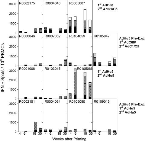
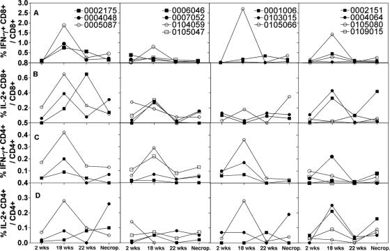
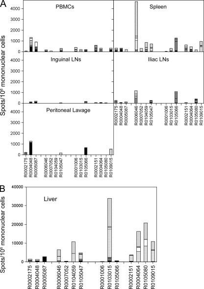
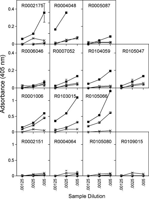
In this study we compared a prime-boost regimen with two serologically distinct replication-defective adenovirus (Ad) vectors derived from chimpanzee serotypes C68 and C1 expressing Gag, Pol, gp140, and Nef of human immunodeficiency virus type 1 with a regimen in which replication-defective Ad vectors of the human serotype 5 (AdHu5) were given twice. Experiments were conducted in rhesus macaques that had or had not been preexposed to antigens of AdHu5. There was no significant difference in T-cell responses tested from peripheral blood of the different groups, although responses were overall highest in nonpreexposed animals immunized with the chimpanzee Ad vectors. Preexisting immunity to AdHu5 completely inhibited induction of transgene product-specific antibodies by the AdHu5 vectors without affecting antibody responses to the chimpanzee vectors. Upon euthanasia, T-cell responses were tested from a number of tissues. Preexisting immunity to AdHu5, commonly found in humans, changed the homing pattern of vaccine-induced T cells. In AdHu5-preexposed animals vaccinated with the chimpanzee Ad vectors, frequencies of transgene-specific T cells were higher in spleens than in blood, and in most preexposed animals vaccinated either with AdHu5 vectors or chimpanzee adenovirus vectors, frequencies of such T cells were exceptionally high in livers. The latter results indicate that analysis of T-cell responses solely from blood mononuclear cells of vaccine recipients may not suffice to compare the potencies of different vaccine regimens.


Figures

FIG. 1.
FIG. 1.
Two groups of four NHPs were injected with 2 × 10¹¹ VP of AdHu5 expressing A1AT. Thirty-four days later, preexposed and nonpreexposed NHPs were vaccinated. Groups 1 and 2 (upper two panels) were immunized with AdC68 vectors expressing Gag, gp140, 5′pol, or TPAnef-3′pol. Animals of groups 3 and 4 (lower two panels) were immunized with the corresponding AdHu5 vectors. The control animal (R0108033) was primed with an AdC68rab.gp vector. T-cell responses from blood were tested 4 and 6 weeks after vaccination by ELISPOT for IFN-γ. NHPs were boosted 120 days later with a second dose of vector. Animals of groups 1 and 2 were boosted with AdC1/C5 vector expressing Gag, gp140, 5′pol, or TPAnef-3′pol. Animals of groups 3 and 4 were boosted with the corresponding AdHu5 vector. The control animal of group 5 was boosted with AdC1/C5rab.gp vector. T-cell responses were tested from blood 2 and 17 weeks after the boost. The graph shows responses of individual animals against seven different pools of peptides. Background data (no peptide) were subtracted. Data for the control animal are not shown. PBMCs from this animal showed the following cumulative spots (i.e., sum of spots obtained with all of the peptide pools minus background spots)/106 cells: week 4, 18; week 6, 0; week 18, 96; week 35, 61. Responses to individual pools were <55 spots/106 PBMCs and thus failed to meet our criteria for a positive response. The different patterns on the bars show responses to different peptide pools. Solid black, Gag peptides; bold stripes, two pools of Pol peptides; dots, Nef peptides; thin stripes and no pattern, three pools of Env peptides.
FIG. 2.
FIG. 2.
The same animals described in Fig. 1 were tested for T-cell responses by ICS of CD3+ CD8+ or CD3+ CD4+ T cells for IFN-γ or IL-2 using 10 pools of peptides. PBMCs were tested 2 weeks after priming, 2 and 6 weeks after the boost, and at the time of necropsy. The background data from the control animal were subtracted. The graphs show the sum of frequencies of CD3+ CD8+ cells secreting IFN-γ (A), CD3+ CD8+ cells secreting IL-2 (B), CD3+ CD4+ cells secreting IFN-γ (C), and CD3+ CD4+ cells secreting IL-2 (D) obtained by adding frequencies obtained with individual peptide pools.
FIG. 3.
FIG. 3.
Lymphocytes from different tissues from the same NHPs described in Fig. 1 were tested at the time of necropsy for T cells producing IFN-γ by ELISPOT. (The codes for the animal numbers and the group designations are shown in Table 2.) The shading of the bars, which indicates the cumulative response to the different pools of peptides, is identical to that in Fig. 1. Data for the control animal (group 5) are not shown. (A) Results for cells from blood, spleens, lymph nodes, and peritoneal lavage. For these tissues the control animal showed the following numbers of spots for all peptide pools/106 mononuclear cells: blood, 54; inguinal lymph nodes, 8; iliac lymph nodes, 36; peritoneal lavage, 46; spleen, 51. All responses to individual pools were <55 spots/106 cells and thus failed to meet our criteria for a positive response. (B) Results for cells isolated from the liver, shown on a different scale. Cells isolated from the liver of the control monkey showed 0 spots per 106 mononuclear cells.
FIG. 4.
FIG. 4.
Sera from the same NHPs shown in Fig. 1 were tested for antibodies to Gag before and after each vaccine dose. The graphs, which are organized in the same fashion as for Fig. 1, show mean absorbance ± standard deviations at three serum dilutions. The Xs indicate sera harvested before vaccination, half-filled squares indicate sera harvested after the first immunization, and filled squares indicate sera harvested after the boost. The control animal did not develop antibodies to Gag (not shown).

References

    1. Casimiro, D. R., L. Chen, T. M. Fu, R. K. Evans, M. J. Caulfield, M. E. Davies, A. Tang, M. Chen, L. Huang, V. Harris, D. C. Freed, K. A. Wilson, S. Dubey, D. M. Zhu, D. Nawrocki, H. Mach, R. Troutman, L. Isopi, D. Williams, W. Hurni, Z. Xu, J. G. Smith, S. Wang, X. Liu, L. Guan, R. Long, W. Trigona, G. J. Heidecker, H. C. Perry, N. Persaud, T. J. Toner, Q. Su, X. Liang, R. Youil, M. Chastain, A. J. Bett, D. B. Volkin, E. A. Emini, and J. W. Shiver. 2003. Comparative immunogenicity in rhesus monkeys of DNA plasmid, recombinant vaccinia virus, and replication-defective adenovirus vectors expressing a human immunodeficiency virus type 1 gag gene. J. Virol. 77:6305-6313. - PMC - PubMed
    1. Cohen, P. 2006. Immunity's yin and yang. A successful vaccine must first avoid being eliminated by pre-existing immunity before it can promote a protective immune response. IAVI Rep. 10:1-5. - PubMed
    1. Crystal, R. G. 1992. Gene therapy strategies for pulmonary disease. Am. J. Med. 92:44S-52S. - PubMed
    1. DiPaolo, N., S. Ni, A. Gaggar, R. Strauss, S. Tuve, Z. Y. Li, D. Stone, D. Shayakhmetov, N. Kiviat, P. Toure, S. Sow, B. Horvat, and A. Lieber. 2006. Evaluation of adenovirus vectors containing serotype 35 fibers for vaccination. Mol. Ther. 13:756-765. - PMC - PubMed
    1. Durier, C., O. Launay, V. Meiffredy, Y. Saidi, D. Salmon, Y. Levy, J. G. Guillet, G. Pialoux, and J. P. Aboulker. 2006. Clinical safety of HIV lipopeptides used as vaccines in healthy volunteers and HIV-infected adults. AIDS 20:1039-1049. - PubMed

Publication types