. 2007 Jun;81(12):6771-4.
doi: 10.1128/JVI.02231-06. Epub 2007 Apr 11.

New structure model for the packaging signal in the genome of group IIa coronaviruses

Affiliations

New structure model for the packaging signal in the genome of group IIa coronaviruses

Shih-Cheng Chen et al. J Virol. 2007 Jun.

Abstract

A 190-nucleotide (nt) packaging signal (PS) located in the 3' end of open reading frame 1b in the mouse hepatitis virus, a group IIa coronavirus, was previously postulated to direct genome RNA packaging. Based on phylogenetic data and structure probing, we have identified a 95-nt hairpin within the 190-nt PS domain which is conserved in all group IIa coronaviruses but not in the severe acute respiratory syndrome coronavirus (group IIb), group I coronaviruses, or group III coronaviruses. The hairpin is composed of six copies of a repeating structural subunit that consists of 2-nt bulges and 5-bp stems. We propose that repeating AA bulges are characteristic features of group IIa PSs.


Figures
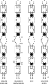
FIG. 1.
FIG. 1.
Highly conserved secondary structure of the PS of group IIa CoVs. Covariations are highlighted in black boxes. The conserved AGC/GUAAU motifs are surrounded with a box of solid lines, while the other motif is surrounded with a box of dashed lines. The GGU insertion in the internal loop of HCoV OC43 and HEV is surrounded by the small stippled box. Residues in parentheses indicate the sequence of closely related species or isolates.
FIG. 2.
FIG. 2.
Structure probing of the MHV PS. (A) Secondary-structure model of the MHV PS. Nucleotides are numbered from the 5′ end of the first cytosine in the PS. Bulges, the internal loop, and the pentaloop are indicated. Additional base pairs in the internal loop are indicated by a dotted line. (B to D) Enzymatic and chemical probing. Susceptible nucleotides were identified by a reference sequencing ladder. For probing reactions, 0.1-μg aliquots of RNA transcripts were treated with nuclease-free water (N), 0.0001 U of RNase T1 (T1), 0.0001 U of RNase V1 (V1), 4 U of RNase S1 (S1), 10 pg of RNase A (Ra), 0.01% (vol/vol) DEPC (P), and 0.05% (vol/vol) DMS (D) for 20 min at room temperature in a total volume of 50 μl. DMS and DEPC probing shown in Fig. 2D were done on ice. The incubation buffer contained 10 mM Tris, 100 mM KCl, 10 mM MgCl2, and, optionally, 10 mM of zinc acetate (indicated by the asterisk above these lanes), pH 6.0. Primer extension reactions were carried out with 0.01 μg of treated transcripts, 0.5 μl of a 0.1 mM concentration of the MHV1 primer, 1 μl of 5 mM dGAT, 1 μl of 25 μM dCTP, 0.1 μl of α-32P-labeled dCTP (10 mCi/ml), 1 μl of 5× reverse transcriptase buffer (Promega), and 20 U of Moloney murine leukemia virus reverse transcriptase (Promega).
FIG. 3.
FIG. 3.
Predicted effects of mutations on the structure of the 69-nt MHV PS. Structure of the wild-type 69-nt MHV PS (A) adapted from the work of Fosmire et al. (3) and (B) redrawn to the new model. The boxed residues indicate the sequences of the FB series mutants FB7, FB8, FB12, and FB13, which were constructed by Fosmire et al. (3), where Δ stands for deletion. The two extra residues (g and c) at the 5′ end were vector derived and stabilized the structure shown in panel B.

References

    1. Altschul, S. F., W. Gish, W. Miller, E. W. Myers, and D. J. Lipman. 1990. Basic local alignment search tool. J. Mol. Biol. 215:403-410. - PubMed
    1. Cologna, R., and B. G. Hogue. 2000. Identification of a bovine coronavirus packaging signal. J. Virol. 74:580-583. - PMC - PubMed
    1. Fosmire, J. A., K. Hwang, and S. Makino. 1992. Identification and characterization of a coronavirus packaging signal. J. Virol. 66:3522-3530. - PMC - PubMed
    1. Gorbalenya, A. E., E. J. Snijder, and W. J. M. Spaan. 2004. Severe acute respiratory syndrome coronavirus phylogeny: toward consensus. J. Virol. 78:7863-7866. - PMC - PubMed
    1. Hsieh, P.-K., S. C. Chang, C.-C. Huang, T.-T. Lee, C.-W. Hsiao, Y.-H. Kou, I.-Y. Chen, C.-K. Chang, T.-H. Huang, and M.-F. Chang. 2005. Assembly of severe acute respiratory syndrome coronavirus RNA packaging signal into virus-like particles is nucleocapsid dependent. J. Virol. 79:13848-13855. - PMC - PubMed

Publication types