. 2007 Jul;81(13):6993-7000.
doi: 10.1128/JVI.00244-07. Epub 2007 Apr 11.

The abundant R2 mRNA generated by aleutian mink disease parvovirus is tricistronic, encoding NS2, VP1, and VP2

Affiliations

The abundant R2 mRNA generated by aleutian mink disease parvovirus is tricistronic, encoding NS2, VP1, and VP2

Jianming Qiu et al. J Virol. 2007 Jul.

Abstract

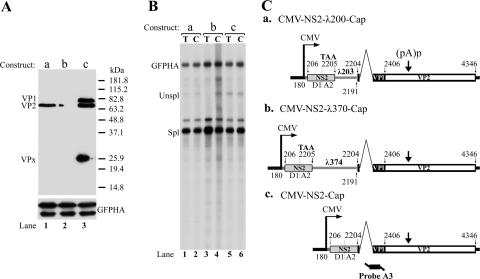
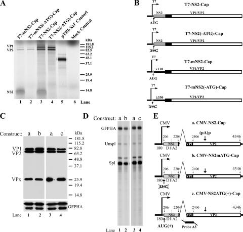
The abundant R2 mRNA encoded by the single left-end promoter of Aleutian mink disease parvovirus is tricistronic; it not only expresses the capsid proteins VP1 and VP2 but is also the major source for the nonstructural protein NS2. A cis-acting sequence within the NS2 gene was shown to be required for efficient capsid protein production, and its effect displayed a distinct location dependence. Ribosome transit through the upstream NS2 gene region was necessary for efficient VP1 and VP2 expression; however, neither ablation nor improvement of the NS2 initiating AUG had an effect on capsid protein production, suggesting that the translation of the NS2 protein per se had little influence on VP1 and VP2 expression. Thus, proper control of the alternative translation of the tricistronic R2 mRNA, a process critical for viral replication, is governed in a complex manner.


Figures

FIG. 1.
FIG. 1.
Genetic map of AMDV-G. The genome of AMDV-G is shown to scale, with the major transcription units indicated, including the inverted hairpin termini (TR), promoters, splice donors (D1, D2, and D3), and acceptors (A1, A2, and A3) as well as the internal proximal and distal polyadenylation sites [(pA)p and (pA)d, respectively]. The AMDV-G proteins that are predicted to be encoded from each RNA are indicated. The sizes of all RNAs, minus polyadenylated tails, are given in parentheses (in nucleotides). Within the nonstructural region, the black bar indicates NS1-encoding ORF1, the striped box indicates NS3-encoding ORF2, and the gray box indicates NS2-encoding ORF3. The large capsid protein-encoding ORF shared by VP1 and VP2 in the right-hand end of the genome is read in ORF3, which is indicated by an open box.
FIG. 2.
FIG. 2.
The R2 mRNA encodes both NS2 and capsid proteins VP1 and VP2. CMV-driven AMDV-G R2-expressing constructs CMV-HA-NS2-Cap and CMV-HA-NS2Cap(pA)p(−), diagrammed in C and designated a and b, respectively, were transfected into CrFK cells. The location of the inserted HA tag is shown. The predicted RNA products that they generate, their relevant ORFs, and their protein products are indicated. Two days following transfection, total cell lysates were resolved on SDS-10% PAGE gels and analyzed by Western blotting using antibodies against either VP1/VP2 (lanes 1 and 2) or the HA-tagged NS2 protein (lanes 3 and 4) (A). An HA-tagged GFP plasmid was cotransfected as a control (lanes 3 and 4). VP1, VP2, GFP, and NS2 protein bands are indicated. The small band at approximately 25 kDa in lanes 1 and 2, which also appears in other figures, marked VPx, is a specific caspase cleavage product of the capsid proteins, which will be described elsewhere (Qiu and Pintel, unpublished). (B) RNase protection analysis of total RNA protected by probe A3 (lanes 1 and 2) and probe P(PA)p (lane 3 and 4). Probes and their protected bands are diagramed at the bottom of C (panel b), with their respective sizes given in nucleotides. Unspl, unspliced; Spl, spliced.
FIG. 3.
FIG. 3.
Substitution or deletion of NS2 reduces VP1 and VP2 expression. The constructs used for this experiment are shown in C and are designated a to d, as are the relevant ORFs used and the potential products generated. The protein products generated by these constructs, as indicated above the lanes, are shown in the Western blot in A. VP1, VP2, β-actin, VPx, and GFP are indicated. (B) RNase protection assays using probe A3 (lanes 1 to 4) and probe P(pA)p (lanes 5 to 8) were performed on cytoplasmic RNA taken from the same experiments, showing that all constructs generate similar amounts of cytoplasmic RNA and a similar ratio of (pA)p to (pA)d. Probes are diagrammed in C. Expression levels of HA-tagged GFP and β-actin (in lanes 1 and 2) were used as an internal control; protein expression is shown at the bottom of A, and RNA accumulation is shown as the GFP-HA band in B. AMDV-G RNA is indicated as unspliced (Unspl), spliced (Spl), RNA polyadenylated at (pA)p [(pA)p], and RNA polyadenylated at (pA)d [(pA)d].
FIG. 4.
FIG. 4.
Effect of the NS2 cis element on the expression of VP1 and VP2 is location dependent. The constructs used, as described in the text, are shown in C and are designated a to c. The relevant ORFs used, the inserted heterologous λ DNA fragments, and an indication of the improved NS2 AUG signal are also shown. The protein products generated by these constructs are shown in the Western blot in A. VP1, VP2, and VPx are indicated. B shows an RNase protection assay of total and cytoplasmic RNA taken from the same experiment using probe A3, which is diagrammed at the bottom of C. Expression of HA-tagged GFP was used as an internal control, protein expression is shown at the bottom of A, and RNA accumulation is shown as the GFP-HA band in B. Unspl, unspliced; Spl, spliced; T, total RNA; C, cytoplasmic RNA.
FIG. 5.
FIG. 5.
R2 mRNA generates NS2 and VP1/VP2 in vitro, and translation of the NS2 ORF does not affect proper capsid protein production. T7-driven constructs used for the generation of mRNA, as described in the text, are shown in B. RNA was transcribed in vitro and was capped and poly(A) tailed. Rabbit reticulocyte lysate was used for in vitro translation in the presence of [35S]methionine. Translated proteins were resolved by SDS-10% PAGE gels (A). The gel was dried and imaged by autoradiography. Individual bands of VP1, VP2, and NS2 are indicated along with their respective sizes. Lane 5 shows a positive in vitro translation control (Xef mRNA; supplied by the manufacturer), and lane 6 is a mock control. CMV-driven R2-expressing construct CMV-NS2-Cap (E, panel a) and the NS2 ATG mutants described in the text (E, panels b and c) were transfected into CrFK cells. Forty-eight hours posttransfection, total cell lysates from transfections using plasmids as indicated were resolved on SDS-10% PAGE gels and analyzed by Western blotting using antibody against the capsid proteins (C). The VP1, VP2, and VPx protein bands are indicated. Cytoplasmic RNA extracted from the same experiment was analyzed by RNase protection assay (D) using probe A3, which is diagrammed in E (panel c). An HA-tagged GFP expression plasmid was cotransfected as a control, and protein expression and RNA expression are indicated (C and D).
FIG. 6.
FIG. 6.
Hairpin structures within the NS2 gene region block VP1 and VP2 expression. The parent plasmid (CMV-NS2-Cap) and mutants described in the text with hairpin insertions in the 5′ UTR (CMV-5×Bam/nt200-NS2-Cap), the beginning of the NS2 second exon (CMV-NS2-5×Bam/nt2059-Cap), the middle of the NS2 first exon (CMV-NS2-5×Bam/nt320-Cap), and the end of the NS2 first exon (CMV-NS2-5×Bam/nt380-Cap) are shown in C and are designated a to e, respectively. A cartoon showing where a ribosome would initiate at the 5′ end of the message is also depicted. The protein products generated following transfection of these constructs are shown in the Western blot in A. VP1, VP2, and VPx are indicated. B shows an RNase protection assay of cytoplasmic RNA taken from the same experiment, using probe A3, which is diagrammed in C (panel e). Expression of HA-tagged GFP was used as an internal control, protein expression is shown at the bottom of A, and cytoplasmic RNA accumulation is shown as the GFP-HA band in B. Unspl, unspliced; Spl, spliced.

Similar articles

Cited by

References

    1. Alexandersen, S. 1986. Acute interstitial pneumonia in mink kits: experimental reproduction of the disease. Vet. Pathol. 23:579-588. - PubMed
    1. Alexandersen, S., M. E. Bloom, and J. Wolfinbarger. 1988. Evidence of restricted viral replication in adult mink infected with Aleutian disease of mink parvovirus. J. Virol. 62:1495-1507. - PMC - PubMed
    1. Alexandersen, S., M. E. Bloom, J. Wolfinbarger, and R. E. Race. 1987. In situ molecular hybridization for detection of Aleutian mink disease parvovirus DNA by using strand-specific probes: identification of target cells for viral replication in cell cultures and in mink kits with virus-induced interstitial pneumonia. J. Virol. 61:2407-2419. - PMC - PubMed
    1. Alexandersen, S., A. Uttenthal-Jensen, and B. Aasted. 1986. Demonstration of non-degraded Aleutian disease virus (ADV) proteins in lung tissue from experimentally infected mink kits. Arch. Virol. 87:127-133. - PubMed
    1. Becerra, S. P., J. A. Rose, M. Hardy, B. M. Baroudy, and C. W. Anderson. 1985. Direct mapping of adeno-associated virus capsid proteins B and C: a possible ACG initiation codon. Proc. Natl. Acad. Sci. USA 82:7919-7923. - PMC - PubMed

Publication types

MeSH terms

LinkOut - more resources