

Human protothecosis
- PMID: 17428884
- PMCID: PMC1865593
- DOI: 10.1128/CMR.00032-06
Human protothecosis
Abstract
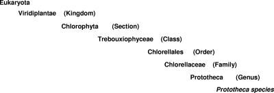
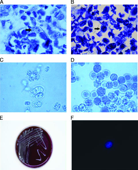
Human protothecosis is a rare infection caused by members of the genus Prototheca. Prototheca species are generally considered to be achlorophyllic algae and are ubiquitous in nature. The occurrence of protothecosis can be local or disseminated and acute or chronic, with the latter being more common. Diseases have been classified as (i) cutaneous lesions, (ii) olecranon bursitis, or (iii) disseminated or systemic manifestations. Infections can occur in both immunocompetent and immunosuppressed patients, although more severe and disseminated infections tend to occur in immunocompromised individuals. Prototheca wickerhamii and Prototheca zopfii have been associated with human disease. Usually, treatment involves medical and surgical approaches; treatment failure is not uncommon. Antifungals such as ketoconazole, itraconazole, fluconazole, and amphotericin B are the most commonly used drugs to date. Among them, amphotericin B displays the best activity against Prototheca spp. Diagnosis is largely made upon detection of characteristic structures observed on histopathologic examination of tissue.
Figures
References
-
- Ahbel, D., A. Alexander, M. Kleine, and D. Lichtman. 1980. Protothecal olecranon bursitis. A case report and review of the literature. J. Bone Joint Surg. 62:835-836. - PubMed
-
- Arnold, P., and D. G. Ahearn. 1972. The systematics of the genus Prototheca with a description of a new species P. filamenta. Mycologia 64:265-275.
-
- Ashford, B., R. Ciferri, and L. Dalmau. 1930. A new species of Prototheca and a variety of the same isolated from the human intestine. Arch. Protistk. 70:619-638.
-
- Begum, F., and P. Syrett. 1970. Fermentation of glucose by Chlorella. Arch. Protistk. 72:344-352. - PubMed
Publication types
MeSH terms
Substances
LinkOut - more resources
Full Text Sources
Miscellaneous
