. 2007 Apr 17;104(16):6776-81.
doi: 10.1073/pnas.0611244104. Epub 2007 Apr 11.

HIV controllers exhibit potent CD8 T cell capacity to suppress HIV infection ex vivo and peculiar cytotoxic T lymphocyte activation phenotype

Affiliations

HIV controllers exhibit potent CD8 T cell capacity to suppress HIV infection ex vivo and peculiar cytotoxic T lymphocyte activation phenotype

Asier Sáez-Cirión et al. Proc Natl Acad Sci U S A. .

Abstract

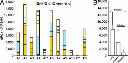
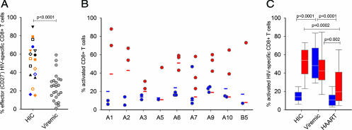
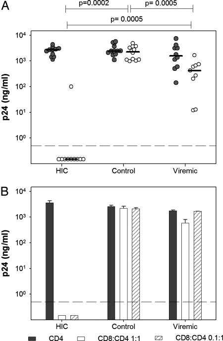
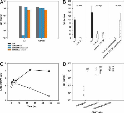
Some rare HIV-1-infected individuals, referred to as HIV controllers (HIC), have persistently undetectable plasma viral load in the absence of therapy. This control of HIV-1 replication has been associated with a strong, multifunctional specific CD8(+) T cell response. However, no direct link between this immune response and the control of viremia has so far been provided. We investigated parameters of specific CD8(+) T cell response and in vitro susceptibility to HIV-1 infection in 11 HIC. We found high frequencies of HIV-specific CD8(+) T cells. Interestingly, these cells expressed the activation marker HLA-DR but not CD38. This unique phenotype differentiates HIV-specific CD8(+) T cells from HIC and noncontroller subjects and likely reflects a high potential to expand upon exposure to antigen and a capacity to exert effector functions. Accordingly, although CD4(+) T cells from HIC were fully susceptible to HIV-1 superinfection, their CD8(+) T cells effectively suppressed HIV-1 infection. Remarkably, this potent anti-HIV activity was observed without prior stimulation of CD8(+) T cells. This activity was not mediated by secreted inhibitory factors but was due to the elimination of infected CD4(+) T cells and was observed only with autologous CD4(+) T cells, indicating an HLA-restricted cytotoxic mechanism. This constitutive antiviral capacity of CD8(+) T cells could account for the control of viral replication in HIC.


Conflict of interest statement

The authors declare no conflict of interest.

Figures

Fig. 1.
Fig. 1.
Frequencies of HIV-specific IFN-γ-secreting CD8+ T cells in HIC, viremic, and HAART subjects. (A) For all subjects (except A5) an average of 41 ± 9 individual peptides were tested, depending on the results of HLA typing. Each bar corresponds to the sum of SFC per 106 PBMC obtained with peptides described to be restricted by HLA-B57 (orange), HLA-B27 (blue), or other HLA antigens (yellow). ∗, For subject A5, whose HLA-typing was lacking at the time of the study, 12 pools of optimal peptides were used instead of individual peptides. The bar for A5 corresponds to the sum of SFC per 106 PBMC obtained with these pools. (B) Comparison of SFC (mean ± SD) in PBMC from HIC, viremic, and HAART subjects. Statistical differences in SFC between groups are indicated above the bars.
Fig. 2.
Fig. 2.
Differentiation and activation status of HIV-specific CD8+ T cells from HIC and comparison with viremic and HAART subjects. (A) Individual HIV-specific CD8+ CD27- T cell frequencies on CD8+ tetramer+ cells (left) in nine HIC (each symbol represents one individual, one to three specificities were tested per subject) and viremic subjects. For HIC, results obtained with B*2705-gag 263–272 and B*5701-gag 162–172 tetramers are represented in blue and orange, respectively. (B) Frequencies of CD38 (blue) and HLA-DR (red) expression on total CD8+ T cells (horizontal bars) and HIV-specific CD8+ T cells (circles) in nine HIC. (C) Comparison of CD38 (blue) and HLA-DR (red) expression (median, 25th–75th and 10th–90th percentiles) on HIV-specific CD8+ T cells in HIC, viremic, and HAART groups. Statistical differences between groups are indicated above the bars.
Fig. 3.
Fig. 3.
Control of HIV-1 infection by ex vivo HIC CD8+ T cells. HIV-1 in vitro infection assays were done with cells from HIC, uninfected donors (control), or HIV viremic individuals [median plasma viral load was 25,350 RNA copies per milliliter (range 7,800–321,000)] (viremic). (A) PHA-activated CD4+ T cells were infected with the replicative HIV-1 BaL in the absence (gray) or presence (white) of autologous unstimulated CD8+ T cells (1:1 ratio). Circles represent the average (n = 3 independent infections) peak p24 values for each studied individual. Horizontal lines indicate median values for each group. Statistical differences in CD8+ T cell-mediated inhibition between groups are indicated above the graph. (B) PHA-activated CD4+ T cells (filled bars) and cocultures of autologous unstimulated CD8+ T cells and PHA-activated CD4+ T cells at ratios of 1:1 (open bars) and 0.1:1 (hatched bars) from five HIC, five uninfected blood donors (control), and nine HIV viremic individuals (viremic) were infected with HIV-1 BaL. One representative experiment (HIC A9) is shown. Bars represent peak levels of p24 in supernatants (mean ± SD, n = 3). Values below the dashed lines were at background level.
Fig. 4.
Fig. 4.
Mechanism of HIC CD8+ T cell-mediated control of HIV-1 infection. (A) CD4+ T cells from HIC and from uninfected donors were infected with replicative HIV-1 BaL. CD4+ T cells cultured alone are shown as a reference (gray bars). Autologous unstimulated CD8+ T cells were added directly to the CD4+ T cell culture (blue solid bars) or to Transwell inserts that were placed in the CD4+ T cell-containing well (blue hatched bars). Autologous PHA-stimulated CD8+ T cells were added to Transwell (orange hatched bars). The ratio of CD8+ to CD4+ T cells was 1:1. One representative experiment with HIC subject A1 and one uninfected control is shown. Results are peak p24 levels. (B) CD4+ T cells from HIC subject A4 were infected with a single-round HIV-1 BaL pseudotype bearing the luciferase gene alone (filled bars) or in coculture (1:1 ratio) with unstimulated autologous CD8+ T cells (open bars). (Left) Results (mean ± SD, n = 3) are the percentage of infection relative to luciferase activity detected in lysates of infected CD4+ T cells 48 h after infection. (Center) At this time point, CD8+ T cells from half the coculture were depleted and viral replication was analyzed 2 days later. (Right) Both the CD8:CD4 coculture (open bar) and the repurified CD4+ T cells (hatched bar) were then rechallenged with the HIV-1 BaL pseudotype, and luciferase activity was measured 48 h later. (C) CD4+ T cells from HIC were infected with a HIV-1 vesicular stomatitis virus glycoprotein pseudotype bearing the GFP reporter gene (filled circles). Forty-eight hours later, autologous unstimulated CD8+ T cells were added to half of the CD4+ T cells at a 1:1 ratio (open circles). At the indicated time points, an aliquot of each cell suspension was labeled with anti-CD4 antibodies and the quantity of double-positive CD4+GFP+ was assessed by flow cytometry. One representative experiment with HIC subject A7 is shown. (D) Unstimulated CD8+ T cells from HIC A1, A3, and B5 (circles) and uninfected blood donors (squares) were cocultured (1:1 ratio) with autologous or heterologous HIC CD4+ T cells (A1:A3, A3:A1, and B5:A7: CD8:CD4 T cell cultures) or uninfected controls CD4+ T cells and infected with replicative HIV-1 BaL. The results (average of three independent infections) show peak p24 levels.

References

    1. Cao Y, Qin L, Zhang L, Safrit J, Ho DD. N Engl J Med. 1995;332:201–208. - PubMed
    1. de Quiros JC, Shupert WL, McNeil AC, Gea-Banacloche JC, Flanigan M, Savage A, Martino L, Weiskopf EE, Imamichi H, Zhang YM, et al. J Virol. 2000;74:2023–2028. - PMC - PubMed
    1. Harrer T, Harrer E, Kalams SA, Elbeik T, Staprans SI, Feinberg MB, Cao Y, Ho DD, Yilma T, Caliendo AM, et al. AIDS Res Hum Retroviruses. 1996;12:585–592. - PubMed
    1. Kloosterboer N, Groeneveld PH, Jansen CA, van der Vorst TJ, Koning F, Winkel CN, Duits AJ, Miedema F, van Baarle D, van Rij RP, et al. Virology. 2005;339:70–80. - PubMed
    1. Lambotte O, Boufassa F, Madec Y, Nguyen A, Goujard C, Meyer L, Rouzioux C, Venet A, Delfraissy JF. Clin Infect Dis. 2005;41:1053–1056. - PubMed

Publication types

MeSH terms