Comparative Study
. 2007 Apr 11;27(15):3937-45.
doi: 10.1523/JNEUROSCI.5281-06.2007.

Amygdala neurons differentially encode motivation and reinforcement

Affiliations
Comparative Study

Amygdala neurons differentially encode motivation and reinforcement

Kay M Tye et al. J Neurosci. .

Abstract

Lesion studies demonstrate that the basolateral amygdala complex (BLA) is important for assigning motivational significance to sensory stimuli, but little is known about how this information is encoded. We used in vivo electrophysiology procedures to investigate how the amygdala encodes motivating and reinforcing properties of cues that induce reinstatement of reward-seeking behavior. Two groups of rats were trained to respond to a sucrose reward. The "paired" group was trained with a reward-predictive cue, whereas the "unpaired" group was trained with a randomly presented cue. Both groups underwent identical extinction and reinstatement procedures during which the reward was withheld. The proportion of neurons that were phasically cue responsive during reinstatement was significantly higher in the paired group (46 of 100) than in the unpaired group (8 of 112). Cues that induce reward-seeking behavior can do so by acting as incentives or reinforcers. Distinct populations of neurons responded to the cue in trials in which the cue acted as an incentive, triggering a motivated reward-seeking state, or as a reinforcer, supporting continued instrumental responding. The incentive motivation-encoding population of neurons (34 of 46 cue-responsive neurons; 74%) extinguished in temporal agreement with a decrease in the rate of instrumental responding. The conditioned reinforcement-encoding population of neurons (12 of 46 cue-responsive neurons; 26%) maintained their response for the duration of cue-reinforced instrumental responding. These data demonstrate that separate populations of cue-responsive neurons in the BLA encode the motivating or reinforcing properties of a cue previously associated with a reward.


Figures
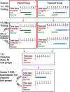
Figure 1.
Figure 1.
Diagram of behavioral paradigm. A cue-induced reinstatement paradigm was modified to allow dissociation of sensory and motor confounds. For the paired group, sucrose delivery was always paired with the LT-cue; for the unpaired group, the LT-cue was presented randomly. However, note that the LT-cue followed 50% of responses for both groups during the reinstatement session. In an FR1 schedule, every response is reinforced. In a random ratio 2 (RR2) schedule, each response has a 0.5 probability of being reinforced.
Figure 2.
Figure 2.
Recording electrode tip placements and example waveforms. A, Coronal diagrams showing electrode tip placements for the paired (red circles) and unpaired (blue circles) groups. Numbers on the left indicate the anteroposterior coordinates caudal to bregma (Paxinos and Watson, 1998). B, Example waveforms of single units recorded from a single electrode tip located in the BLA and isolated using principle component cluster analysis.
Figure 3.
Figure 3.
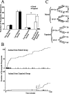
Behavioral differences between paired and unpaired groups. A, Mean numbers of responses (± SEM) for paired (n = 5) and unpaired (n = 5) groups were not different during training sessions and extinction, but only paired animals showed a cue-induced reinstatement of responding. Data analyzed with ANOVA revealed the main effects of group (F(1,16) = 16.23; p < 0.001), session (F(1,16) = 6.57; p < 0.03), and a group × session interaction (F(1,16) = 13.36; p < 0.003) and were followed by pairwise comparisons (Holm-Sidak). *p < 0.001. B, Cumulative activity records depicting the response pattern for representative animals of each group during the 120 min reinstatement session. Each upward inflection and corresponding tick mark represents one nosepoke response, shown on the y-axis. Note slope change for the paired animal after the first ∼65 nosepoke responses. Arrows represent noncontingent sucrose deliveries for the unpaired group 80 min into the reinstatement session. C, Microanalysis of behavior during the reinstatement session. Mean (± SEM) percentages of port entries or nosepokes are given. In the 2 s after the nosepoke, paired animals were more likely to enter the port after a cue (paired t test; p < 0.005) and to repeat responding after the absence of a cue (p < 0.003), whereas unpaired animals were equally likely to enter the port or to repeat nosepoking after either the presence or absence of the cue (p > 0.05). NP, nosepoke; Ø, absence of cue; PE, port entry. *p < 0.005.
Figure 4.
Figure 4.
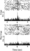
Example of a selectively cue-responsive neuron. The perievent raster and histogram (PERH) of a neuron showed a significant change in phasic activity in response to nosepokes paired with a cue presentation but did not show a significant change in phasic activity to nosepokes that were not paired with the cue. Neurons showing this response pattern are referred to as selectively cue responsive. For all PERHs, the arrow indicates the time of the reference event, or zero time point. The bin size equals 40 ms for this and subsequent figures.
Figure 5.
Figure 5.
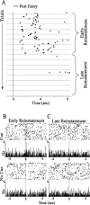
Corresponding changes in behavior and neural encoding of the cue from early to late reinstatement. A, Example of the pattern of port entry during the reinstatement session for a representative paired animal shown in a perievent raster with response-contingent cue presentation as the reference event. Each row represents a trial, and the cue presentation is the reference event. Early in the session, cue presentations were consistently followed by port entries (filled diamonds). A discrete change occurred in late reinstatement because the paired animal continued to respond to the cue but no longer checked the port for sucrose. B, C, Perievent raster and histogram (PERH) of a BLA single-unit with a differential response to the presence (excitation) and absence (inhibition) of the cue in early reinstatement (B) and then a loss of these responses in late reinstatement (C).
Figure 6.
Figure 6.
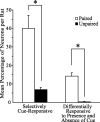
Neural responses to cue presentation. Two types of cue-responsive neurons include selectively cue-responsive neurons and neurons that were differentially responsive to the presence and absence of the cue. The paired group had significantly higher mean percentages of neurons per rat selectively responding to the cue, as well as mean percentages of neurons differentially responding to the presence or absence of the cue, than the unpaired group (n = 5 rats per group; *p < 0.001; t test).
Figure 7.
Figure 7.
Cue-responsive neurons can be categorized into rapidly extinguishing or long-lasting response types. A, Perievent raster and histogram (PERH) of BLA single-unit encoding conditioned incentive; the phasic response to the cue is present only when the subject entered the port after cue presentation. Once the cue no longer provided an incentive to approach the sucrose port, the response to the cue disappeared. B, PERH of BLA single-unit encoding conditioned reinforcement; the phasic response to the cue is present whether or not the subject entered the port after cue presentation. The cue acted as a conditioned reinforcer throughout the reinstatement session, as shown by the ability of the cue presentation to maintain a steady rate of nosepoke responding in the absence of the primary reinforcer.
Figure 8.
Figure 8.
Subpopulations of cue-responsive neurons. The distribution of numbers of cue-responsive neurons recorded from animals in the paired group that are conditioned incentive encoding or conditioned reinforcement encoding.

References

    1. Arroyo M, Markou A, Robbins TW, Everitt BJ. Acquisition, maintenance and reinstatement of intravenous cocaine self-administration under a second-order schedule of reinforcement in rats: effects of conditioned cues and continuous access to cocaine. Psychopharmacology (Berl) 1998;140:331–344. - PubMed
    1. Balleine BW, Killcross AS, Dickinson A. The effect of lesions of the basolateral amygdala on instrumental conditioning. J Neurosci. 2003;23:666–675. - PMC - PubMed
    1. Baxter MG, Parker A, Lindner CC, Izquierdo AD, Murray EA. Control of response selection by reinforcer value requires interaction of amygdala and orbital prefrontal cortex. J Neurosci. 2000;20:4311–4319. - PMC - PubMed
    1. Berridge KC, Robinson TE. Parsing reward. Trends Neurosci. 2003;26:507–513. - PubMed
    1. Cador M, Robbins TW, Everitt BJ. Involvement of the amygdala in stimulus-reward associations: interaction with the ventral striatum. Neuroscience. 1989;30:77–86. - PubMed

Publication types