

Accelerated brain gray matter loss in fibromyalgia patients: premature aging of the brain?
- PMID: 17428976
- PMCID: PMC6672521
- DOI: 10.1523/JNEUROSCI.0098-07.2007
Accelerated brain gray matter loss in fibromyalgia patients: premature aging of the brain?
Abstract
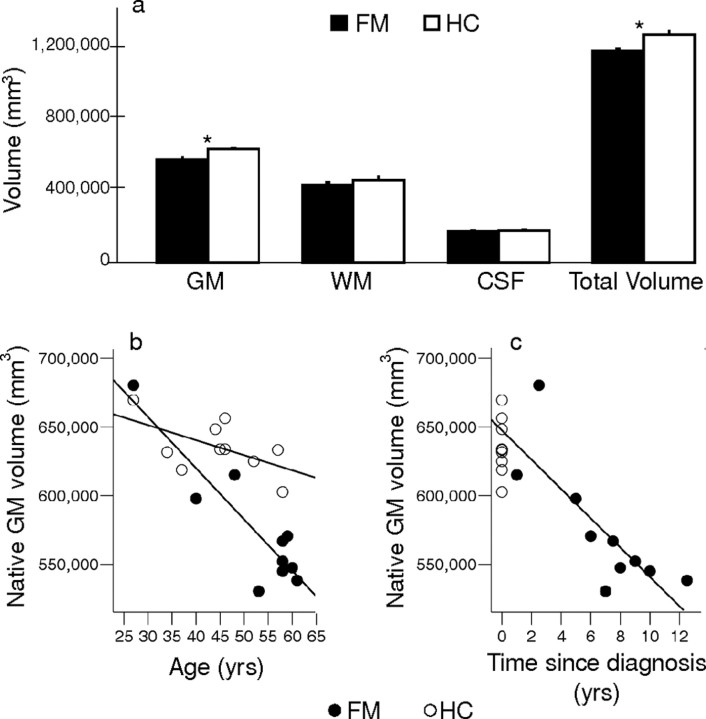
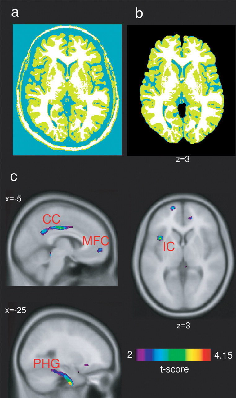
Fibromyalgia is an intractable widespread pain disorder that is most frequently diagnosed in women. It has traditionally been classified as either a musculoskeletal disease or a psychological disorder. Accumulating evidence now suggests that fibromyalgia may be associated with CNS dysfunction. In this study, we investigate anatomical changes in the brain associated with fibromyalgia. Using voxel-based morphometric analysis of magnetic resonance brain images, we examined the brains of 10 female fibromyalgia patients and 10 healthy controls. We found that fibromyalgia patients had significantly less total gray matter volume and showed a 3.3 times greater age-associated decrease in gray matter than healthy controls. The longer the individuals had had fibromyalgia, the greater the gray matter loss, with each year of fibromyalgia being equivalent to 9.5 times the loss in normal aging. In addition, fibromyalgia patients demonstrated significantly less gray matter density than healthy controls in several brain regions, including the cingulate, insular and medial frontal cortices, and parahippocampal gyri. The neuroanatomical changes that we see in fibromyalgia patients contribute additional evidence of CNS involvement in fibromyalgia. In particular, fibromyalgia appears to be associated with an acceleration of age-related changes in the very substance of the brain. Moreover, the regions in which we demonstrate objective changes may be functionally linked to core features of the disorder including affective disturbances and chronic widespread pain.
Figures
References
-
- Apkarian AV, Bushnell MC, Treede RD, Zubieta JK. Human brain mechanisms of pain perception and regulation in health and disease. Eur J Pain. 2005;9:463–484. - PubMed
-
- Chen S, Xia W, Li L, Liu J, He Z, Zhang Z, Yan L, Zhang J, Hu D. Gray matter density reduction in the insula in fire survivors with posttraumatic stress disorder: a voxel-based morphometric study. Psychiatry Res. 2006;146:65–72. - PubMed
-
- Collins DL, Neelin P, Peters TM, Evans AC. Automatic 3D intersubject registration of MR volumetric data in standardized Talairach space. J Comput Assist Tomogr. 1994;18:192–205. - PubMed
-
- Corbo V, Clement MH, Armony JL, Pruessner JC, Brunet A. Size versus shape differences: contrasting voxel-based and volumetric analyses of the anterior cingulate cortex in individuals with acute posttraumatic stress disorder. Biol Psychiatry. 2005;58:119–124. - PubMed
Publication types
MeSH terms
LinkOut - more resources
Full Text Sources
Other Literature Sources
Medical