Comparative Study
. 2007 Apr 11;27(15):4061-71.
doi: 10.1523/JNEUROSCI.0068-07.2007.

Muscarinic control of long-range GABAergic inhibition within the rhinal cortices

Affiliations
Comparative Study

Muscarinic control of long-range GABAergic inhibition within the rhinal cortices

John Apergis-Schoute et al. J Neurosci. .

Abstract

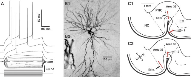
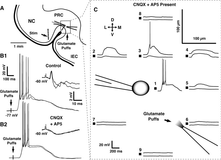
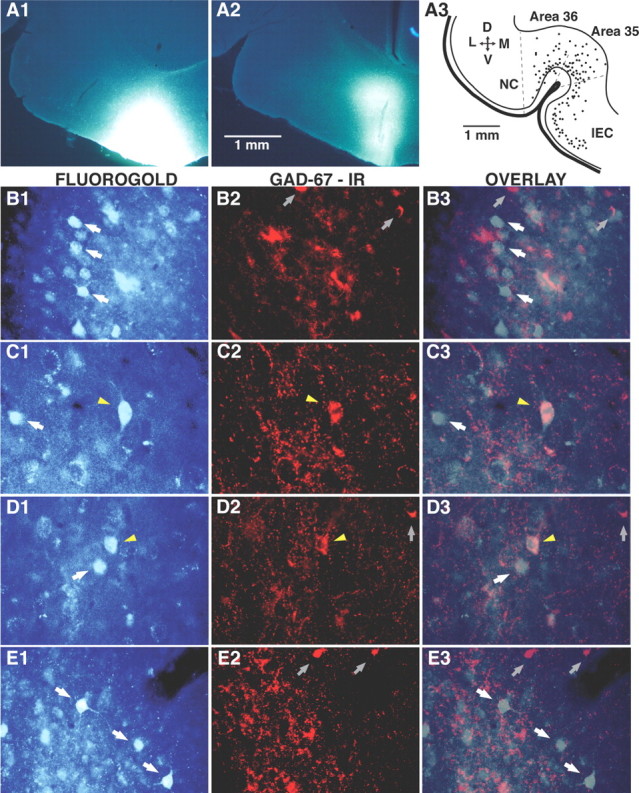
The perirhinal cortex plays a critical role in memory formation, in part because it forms reciprocal connections with the neocortex and entorhinal cortex and is thus in a position to integrate and transfer higher-order information to and from the hippocampus. However, for reasons that remain unclear, perirhinal transfer of neocortical inputs to the entorhinal cortex occurs with a low probability. Using patch recordings in vitro and tract-tracing combined with GAD-67 immunohistochemistry, we show that the perirhinal cortex contains GABAergic neurons with long-range projections to superficial entorhinal cells. This finding challenges the traditional model of cortical inhibition in which all trans-areal inhibition is thought to be disynaptic because the axons of GABAergic interneurons are assumed to be confined within the area in which their somata are located. Moreover, consistent with recent studies indicating that the formation of perirhinal-dependent memories requires activation of muscarinic receptors, long-range IPSPs were presynaptically inhibited by M2 receptor activation. Overall, these results suggest that long-range feedforward inhibition regulates perirhinal transfer of neocortical inputs to the entorhinal cortex, but that cholinergic inputs can presynaptically adjust the impact of this control mechanism as a function of environmental contingencies.


Figures

Figure 1.
Figure 1.
Identification of recorded cells and experimental setup. Physiological (A) and morphological (B) identification of principal rhinal neurons. Whole-cell patch recordings of perirhinal or entorhinal neurons were obtained under visual guidance, and their electroresponsive properties were studied by injecting a series of rectangular current pulses of gradually increasing amplitude. We only considered rhinal neurons that displayed a regular spiking pattern, as shown in the representative example of layer II entorhinal neurons depicted in A. Intracellular Neurobiotin injection was performed to reveal the morphology of recorded cells. As shown in B, all recovered neurons had multipolar dendritic trees (B1) that were covered with spines (B2). C, Experimental setup. A stimulating electrode array (stim.; red dots) was positioned in area 35 (C1) or area 36 (C2), and evoked responses were monitored in cells of the vlEC (C1) or area 35 (C2), respectively. The cross indicates orientation of the scheme in which D, V, L, and M stand for dorsal, ventral, lateral, and medial, respectively. NC, Neocortex.
Figure 2.
Figure 2.
Threshold electrical stimulation of area 35 often elicits CNQX- and AP-5-resistant IPSPs in vlEC neurons. A1, Response of a superficial vlEC neuron to area 35 stimuli of gradually increasing intensity (numbers on the right). A2, Response seen in A1 with 200 μA stimulus is not abolished by addition of CNQX and AP-5 to the aCSF. B, In many vlEC cells, IPSPs preceded EPSPs after threshold stimulation of area 35. B1, Example of IPSP–EPSP sequence evoked from area 35 at three different membrane potentials (numbers on the right), as determined by intracellular current injection. B2, The initial IPSP persisted after addition of CNQX and AP-5 to the aCSF. B3, CNQX- and AP-5-resistant IPSPs evoked in vlEC cells by threshold stimulation of area 35 reverse just below −70 mV (top) and are abolished by picrotoxin (bottom).
Figure 3.
Figure 3.
Threshold electrical stimulation of area 36 often elicits CNQX- and AP-5-resistant IPSPs in area 35 neurons. A, Response of a superficial area 35 neuron to area 36 stimuli of gradually increasing intensity (numbers on the right). B, Response seen in A with 200 μA stimulus is not abolished by addition of CNQX and AP-5 to the aCSF. C, CNQX- and AP-5-resistant IPSPs evoked in area 35 cells by threshold stimulation of area 36 reverse just below −70 mV (top) and are abolished by picrotoxin (bottom).
Figure 4.
Figure 4.
Contrasting effects of CNQX and AP-5 on synaptically released versus pressure-applied glutamate. Control experiments designed to test the feasibility and spatial selectivity of local pressure glutamate applications in the presence of glutamate receptor antagonists. A, Scheme showing the experimental approach used in these control experiments. An array of stimulating electrodes (dots) was positioned in area 36 while monitoring evoked responses with the patch method in superficial area 35 neurons. A second pipette containing 0.5 mm glutamate (dissolved in aCSF) was positioned at proximity of the recorded neuron. B, Responses to pressure-applied (left) or synaptically released (right) glutamate in control aCSF (B1) versus after addition of CNQX and AP-5 to the aCSF (B2). C, Spatial selectivity of responses to pressure-applied glutamate. Pressure applications of glutamate were performed at various locations (100 μm grid pattern; red squares) around a perirhinal cell recorded in whole-cell mode. Two stimulus durations (60 and 100 ms) were tested (bottom and top traces, respectively). Only two of the ejection sites (C1, C3), both located in the immediate vicinity of the soma, evoked suprathreshold responses in this perirhinal neuron. D, Dorsal; V, ventral; L, lateral; M, medial; NC, neocortex.
Figure 5.
Figure 5.
Local pressure application of glutamate in area 35 elicits CNQX- and AP-5-resistant IPSPs in superficial vlEC neurons. A, Response of a layer II vlEC neuron to local pressure application of glutamate in area 35. The same test was performed at three different membrane potentials (numbers on the right), as determined by intracellular current injection. B, In the same neuron, CNQX and AP-5 block the EPSP but not the IPSP elicited by threshold electrical stimulation of area 35. C, A second example of long-range IPSPs evoked in a vlEC neurons by pressure application of glutamate in area 35 in the presence of CNQX and AP-5. Various pulses durations (numbers on the left) were tested.
Figure 6.
Figure 6.
Iontophoretic injection of Fluorogold in the vlEC retrogradely labels perirhinal neurons, a proportion of which are immunoreactive for GAD-67. A1, A2, Representative examples of Fluorogold injection sites in the vlEC. A3, Mapping of retrogradely labeled neurons (dots) in the perirhinal cortex on a representative 60-μm-thick coronal section. B–E are all organized in the same manner. The left column shows neurons retrogradely labeled with Fluorogold. The center column shows GAD-67 immunoreactivity (IR) in the same field. The right column overlays the Fluorogold and GAD-67 labeling. The photomicrographs shown in B and C were obtained in area 35, those in D were obtained in area 36, and those in E were obtained in the lateral amygdala. White arrows indicate FG-positive neurons that were immunonegative for GAD-67. Yellow arrowheads mark FG-positive neurons that were immunoreactive for GAD-67. Gray arrows point to neurons immunoreactive for GAD-67 only. D, Dorsal; V, ventral; L, lateral; M, medial; NC, neocortex.
Figure 7.
Figure 7.
Inhibition of long-range IPSCs by M2 receptor activation. CNQX, AP-5, and the M1 receptor antagonist pirenzepine are present in the aCSF throughout. A, Voltage-clamp recordings of long-range IPSCs evoked in three different layer II vlEC cells by threshold stimulation of area 35. A1, Effect of muscarine (5 μm) on long-range IPSCs. Left, Control response. Right, After addition of muscarine to the aCSF (A2), application of the M2 antagonist methoctramine (0.8 μm) before muscarine completely blocks the inhibition of long-range IPSCs. Left, Control response in the presence of methoctramine. Right, Response elicited in the presence of methoctramine and muscarine. A3, The muscarinic inhibition of long-range IPSCs is also observed when vlEC cells are recorded with pipettes containing a CS+-based internal solution. Left, Control response. Right, Response elicited in the presence of muscarine. B, Muscarine produces an increase in paired-pulse ratio. B1, Top, Control conditions. Bottom, After addition of muscarine. B2, Same IPSCs as in B1 after scaling of the first IPSC seen in the presence of muscarine to that observed in control conditions. C, Graph plotting the ratio of experimental to control 1/CV2 (y-axis) against the ratio of experimental to control response amplitudes (x-axis). The solid line is a linear fit to the data.

References

    1. Abe H, Ishida Y, Iwasaki T. Perirhinal N-methyl-d-aspartate and muscarinic systems participate in object recognition in rats. Neurosci Lett. 2004;356:191–194. - PubMed
    1. Aggleton JP, Hunt PR, Rawlins JN. The effects of hippocampal lesions upon spatial and non-spatial tests of working memory. Behav Brain Res. 1986;19:133–146. - PubMed
    1. Alonso A. Electrophysiology of neurones in the perirhinal and entorhinal cortices and neuromodulatory changes in firing patterns. In: Witter MP, Wouterlood F, editors. The parahippocampal region. Oxford: Oxford UP; 2002. pp. 89–105.
    1. Behrends JC, ten Bruggencate G. Cholinergic modulation of synaptic inhibition in the guinea pig hippocampus in vitro: excitation of GABAergic interneurons and inhibition of GABA-release. J Neurophysiol. 1993;69:626–629. - PubMed
    1. Bekkers JM, Stevens CF. Presynaptic mechanism for long-term potentiation in the hippocampus. Nature. 1990;346:724–729. - PubMed

Publication types

MeSH terms

LinkOut - more resources