Comparative Study
. 2007 Apr 11;27(15):4101-9.
doi: 10.1523/JNEUROSCI.4295-06.2007.

Extensive turnover of dendritic spines and vascular remodeling in cortical tissues recovering from stroke

Affiliations
Comparative Study

Extensive turnover of dendritic spines and vascular remodeling in cortical tissues recovering from stroke

Craig E Brown et al. J Neurosci. .

Abstract

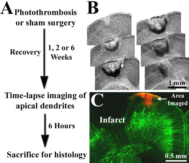
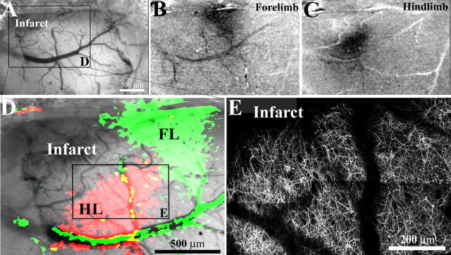
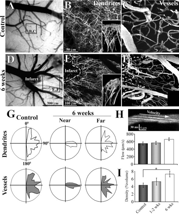
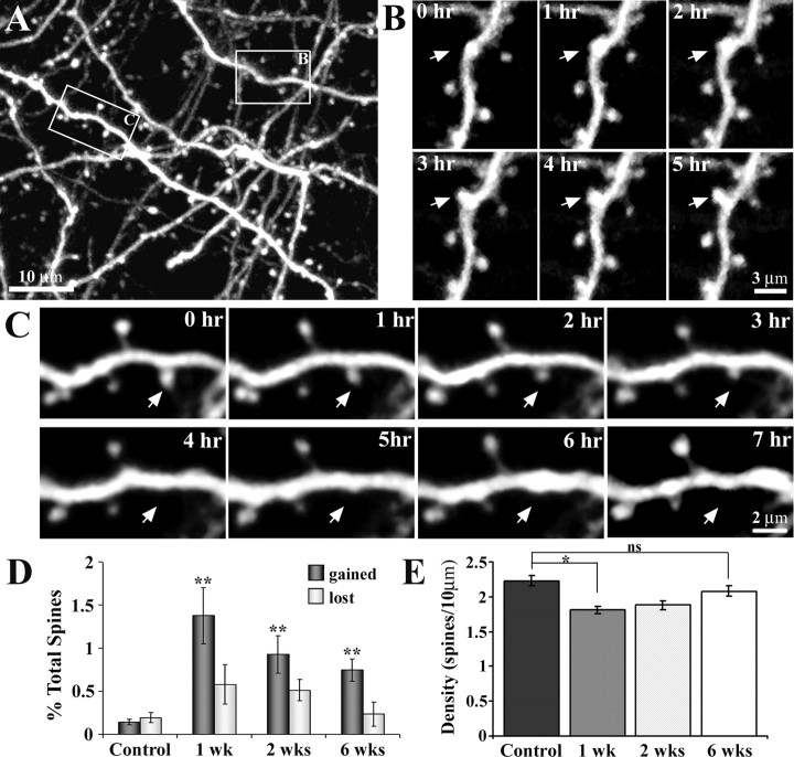
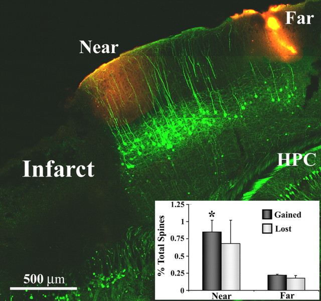
Recovery of function after stroke is thought to be dependent on the reorganization of adjacent, surviving areas of the brain. Macroscopic imaging studies (functional magnetic resonance imaging, optical imaging) have shown that peri-infarct regions adopt new functional roles to compensate for damage caused by stroke. To better understand the process by which these regions reorganize, we used in vivo two-photon imaging to examine changes in dendritic and vascular structure in cortical regions recovering from stroke. In adult control mice, dendritic arbors were relatively stable with very low levels of spine turnover (<0.5% turnover over 6 h). After stroke, however, the organization of dendritic arbors in peri-infarct cortex was fundamentally altered with both apical dendrites and blood vessels radiating in parallel from the lesion. On a finer scale, peri-infarct dendrites were exceptionally plastic, manifested by a dramatic increase in the rate of spine formation that was maximal at 1-2 weeks (5-8-fold increase), and still evident 6 weeks after stroke. These changes were selective given that turnover rates were not significantly altered in ipsilateral cortical regions more distant to the lesion (>1.5 mm). These data provide a structural framework for understanding functional and behavioral changes that accompany brain injury and suggest new targets that could be exploited by future therapies to rebuild and rewire neuronal circuits lost to stroke.


Figures

Figure 1.
Figure 1.
A, Summary of experimental design. B, Representative example of cortical infarction induced by photothrombosis 1 week earlier. Nissl-stained coronal sections, ∼300 μm apart, are arranged in an anterior–posterior manner. In most cases, the infarct zone was centered over the forelimb representation of the sensorimotor cortex and spanned all cortical layers, leaving the white matter intact. C, Coronal section showing the expression of YFP labeling in the cortex. Note that labeling is confined primarily to layer 5 neurons (green), which possess a long apical dendrite that extends to the pial surface and branches laterally. These apical dendritic tufts in peri-infarct cortex were repeatedly imaged to assess dendritic spine turnover and were subsequently marked by DiI injection (red).
Figure 2.
Figure 2.
Dendritic structure and function in peri-infarct cortex. A, Low-magnification brightfield image of the brain's surface in a YFP-transgenic mouse, two weeks after photothrombotic injury. In this case, the cortical infarction (top left in cranial window) occurred just medial and anterior of the forelimb (FL) and hindlimb (HL) representation of the sensorimotor cortex. B, C, To assess whether peri-infarct cortex was viable, functionally responsive cortex, we imaged intrinsic optical signals after stimulating the contralateral forelimb and hindlimb. After somatic stimulation, certain parts of the cortex appear darker, which is indicative of regionally elevated levels of deoxyhemoglobin (which absorbs the light) as a result of increased neuronal activity (Grinvald et al., 1986). The darker regions were in register with stereotaxic coordinates for forelimb and hindlimb regions. D, Enlarged view of boxed area in A showing cortical areas responsive to forelimb (green) and hindlimb (red) stimulation, superimposed over a brightfield image of the cortical surface. E, To compare the borders of functionally responsive tissue with cortical dendritic structure, the apical dendrites in hindlimb cortex (boxed area in D) were imaged in vivo and tiled together (z projections consisting of 50 planar images, 2 μm apart). As shown in D and E, the border between functionally responsive and nonresponsive cortical tissue corresponds well with the infarct border revealed by YFP-labeled dendrites.
Figure 3.
Figure 3.
Reorganization of apical dendritic tufts and flowing vasculature in peri-infarct cortex. A, D, Low-magnification brightfield images showing the surface of the brain and vessels through the cranial window. B, C, E, F, Maximal intensity z projections of 80 planar sections (taken 2 μm apart) illustrating the organization of dendritic tufts and vasculature in a control (B, C) and 6 weeks after photothrombotic stroke (E, F). Insets in B and E show a side view of the apical dendritic tufts projected in the y–z axis. G, Polar plots showing the orientation of dendritic and vascular segments in control (n = 3) and regions near and far from the infarct border (n = 3; near, <250 μm; far, 500–800 μm). Note that 6 weeks after stroke, dendrites and vessels run in parallel with each other at a 90–100 angle in regions close to, but not further away from the infarct border. H, A sample image showing the arc created by flowing Texas Red labeled plasma (offset by negatively stained red blood cells) in the lumen of the capillary. Using the slope of these arcs to assess blood velocity, we found no significant differences in capillary blood flow velocity between control and peri-infarct regions. I, Graph showing a progressive increase in blood vessel density in peri-infarct cortex over time. *p < 0.05.
Figure 4.
Figure 4.
Time-lapse imaging of apical dendrites reveals heightened levels of spine formation in peri-infarct cortex. A, Maximal intensity z projection of 30 planar images (taken 1 μm apart, at 0 h) depicting the Golgi-like detail of YFP-labeled dendrites. In this field of view, a dendritic spine formed (box labeled B) and another retracted (box labeled C) over the imaging period. B, C, Time-lapse images (taken 1 h apart) showing the formation and retraction, respectively, of a dendritic spine. D, Relative to controls, the rate of spine formation was significantly increased in peri-infarct cortex at 1, 2, and 6 weeks after stroke. Spine elimination, although elevated, was not significantly different from control levels. E, Relative to controls, spine density was significantly reduced at 1 and 2 weeks, but appeared to recover by 6 weeks. *p < 0.05; **p < 0.005.
Figure 5.
Figure 5.
Dendritic spine formation is elevated specifically within peri-infarct areas. Representative example of a fixed brain section from a YFP-transgenic mouse (sagittal plane, 2 weeks after stroke) where spine turnover was assessed near and far from the infarct border (sites demarcated with DiI, red-orange color). Inset, Graph showing increased levels of spine formation in regions proximal, but not distal to the infarct. *p < 0.05. HPC, Hippocampus.

References

    1. Biernaskie J, Chernenko G, Corbett D. Efficacy of rehabilitative experience declines with time after focal ischemic brain injury. J Neurosci. 2004;24:1245–1254. - PMC - PubMed
    1. Biernaskie J, Szymanska A, Windle V, Corbett D. Bi-hemispheric contribution to functional motor recovery of the affected forelimb following focal ischemic brain injury in rats. Eur J Neurosci. 2005;21:989–999. - PubMed
    1. Carmeliet P, Tessier-Lavigne M. Common mechanisms of nerve and blood vessel wiring. Nature. 2005;436:193–200. - PubMed
    1. Carmichael ST. Plasticity of cortical projections after stroke. Neuroscientist. 2003a;9:64–75. - PubMed
    1. Carmichael ST. Gene expression changes after focal stroke, traumatic brain and spinal cord injuries. Curr Opin Neurol. 2003b;16:699–704. - PubMed

Publication types