. 2007 Jun;18(6):2313-21.
doi: 10.1091/mbc.e06-11-1040. Epub 2007 Apr 11.

Cell polarity development and protein trafficking in hepatocytes lacking E-cadherin/beta-catenin-based adherens junctions

Affiliations

Cell polarity development and protein trafficking in hepatocytes lacking E-cadherin/beta-catenin-based adherens junctions

Delphine Théard et al. Mol Biol Cell. 2007 Jun.

Abstract

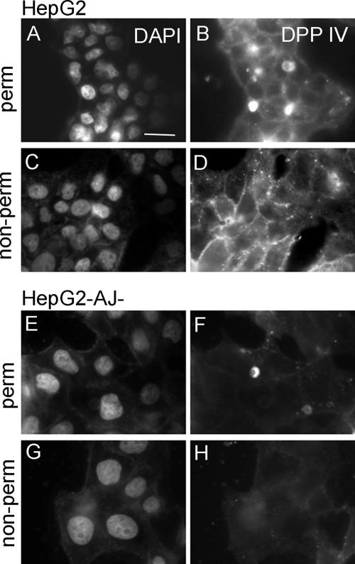
Using a mutant hepatocyte cell line in which E-cadherin and beta-catenin are completely depleted from the cell surface, and, consequently, fail to form adherens junctions, we have investigated adherens junction requirement for apical-basolateral polarity development and polarized membrane trafficking. It is shown that these hepatocytes retain the capacity to form functional tight junctions, develop full apical-basolateral cell polarity, and assemble a subapical cortical F-actin network, although with a noted delay and a defect in subsequent apical lumen remodeling. Interestingly, whereas hepatocytes typically target the plasma membrane protein dipeptidyl peptidase IV first to the basolateral surface, followed by its transcytosis to the apical domain, hepatocytes lacking E-cadherin-based adherens junctions target dipeptidyl peptidase IV directly to the apical surface. Basolateral surface-directed transport of other proteins or lipids tested was not visibly affected in hepatocytes lacking E-cadherin-based adherens junctions. Together, our data show that E-cadherin/beta-catenin-based adherens junctions are dispensable for tight junction formation and apical lumen biogenesis but not for apical lumen remodeling. In addition, we suggest a possible requirement for E-cadherin/beta-catenin-based adherens junctions with regard to the indirect apical trafficking of specific proteins in hepatocytes.


Figures

Figure 1.
Figure 1.
The HepG2-AJ cell line presents a defect in E-cadherin and β-catenin localization. The parental HepG2 and AJ-defective HepG2-AJ cell lines were grown for 72 h, fixed, and immunolabeled for β-catenin (A) and E-cadherin (B). Arrows highlight the presence of β-catenin (A) and E-cadherin (B) at the cell–cell contact in parental HepG2 cells, whereas these proteins are retained intracellularly in HepG2-AJ cells. Bar, 20 μm.
Figure 2.
Figure 2.
The HepG2-AJ cell line develops a polarized phenotype. (A and B) 1-μm-thick coupe from embedded HepG2-AJ cells observed with light microscopy (A) and with transmission electron microscopy (B). Note the presence of intercellular microvilli-lined apical lumens (white arrows in A) despite a clearly perturbed cell–cell contact (cell boundaries are depicted by dashed lines). (C) The localization of different known resident apical proteins (radixin, 5′nucleotidase, and MDR1) is restricted to the plasma membranes lining the intercellular lumens (arrows) in HepG2-AJ cells (corresponding phase contrast images are depicted in the top panels). Bars, 5 μm (A and C), and 2.5 μm (B).
Figure 3.
Figure 3.
Apical lumens occur intracellularly before their exposure at the extracellular space in HepG2-AJ cells. HepG2 and HepG2-AJ cells were plated and fixed at different time points. Apical lumen structures (arrows) were detected with light microscopy and categorized as intracellular (VAC), docked, or intercellular (BC) (see schematic representation in A). (B) Apical lumen structures in 48-h-old HepG2 cells were predominantly intercellular, whereas, by contrast, many apical lumen structures were intracellular in 48-h-old HepG2-AJ cells. (C) Quantification of the distribution of apical lumen structures that were immunolabeled for the microvilli protein villin (mean ± SD of at least 500 cells).
Figure 4.
Figure 4.
Tight junctions are present and functional in HepG2-AJ cells. The localization of the tight junction proteins ZO-1(A, arrow) and PAR3 (B, arrow) was assayed in 3-d old HepG2-AJ cells. Corresponding phase contrast images are shown in the top panels. Bars, 20 μm. (C) HepG2-AJ cells were observed in electron microscopy to detect the presence of tight junctions (arrows). Enlargement in the bottom panel clearly shows TJs and a desmosome (D). Bars, 500 nm (top) and 60 nm (bottom). (D) The gate function (see text) of the tight junctions was examined using CFDA, a fluorescent substrate of MRP2. The HepG2-AJ cells were loaded 30 min at 37°C with 0.5 μM CFDA. Note the restriction of CFDA in the BC lumens (arrows). The corresponding phase-contrast image is depicted in the top panel. Bar, 20 μm. (E) To confirm the proper gate function of the TJ, HepG2-AJ cells were incubated for 30 min at 4°C in presence of 100 μM lucifer yellow. Note that lucifer yellow does not have access to the BC lumens (arrows). The corresponding phase contrast image is depicted in the top panel. Bar, 10 μm. (F and G) The fence function (see text) of the tight junctions was tested using the fluorescent lipid C6-NBD-sphingomyelin. When incorporated in the basolateral plasma membrane at 4°C, the NBD-SM remains at the basolateral membrane, and no lateral diffusion to the apical domain is observed (F, 0 min vs. 15 min). Alternatively, when the fluorescent lipid is loaded into the exoplasmic leaflet of the BC membrane according to a previously described protocol (van IJzendoorn et al., 1997; see Materials and Methods), no lateral diffusion of the lipid probe to the basolateral domain is observed (G, 0 min vs. 15 min). Arrows indicate the localization of the BCs. Bars, 20 μm.
Figure 5.
Figure 5.
HepG2-AJ cells present a defect in bile canalicular lumen remodeling. Parental HepG2 and HepG2-AJ cells were cultured for 5 d, fixed, and stained against phalloidin-tetramethylrhodamine B isothiocyanate (TRITC) to visualize the subapical F-actin meshwork (top). Nuclei are stained with Hoechst (bottom). Note the appearance of extensive multicellular apical lumens in parental HepG2 cells (cf. Herrema et al., 2006), whereas in HepG2-AJ cells apical lumens mainly remain between two adjacent cells, and no apical lumen remodeling has occurred. Bar, 20 μm.
Figure 6.
Figure 6.
E-cadherin blocking antibodies do not prevent the development of polarity in parental HepG2 cells. Parental HepG2 cells were cultured for 3 d in presence of E-cadherin blocking antibody. Cells were then fixed and stained for E-cadherin (A), β-catenin (B, enlargement shows detailed localization of the protein at the membrane), the TJ protein ZO-1 (C), the apical marker radixin (D), and the actin marker phalloidin-TRITC (E). The corresponding phase-contrast pictures are depicted. The arrows in C–E point to the apical lumens. Bar, 20 μm.
Figure 7.
Figure 7.
Steady-state distribution of DPPIV in HepG2 and HepG2-AJ cells. Three-day-old HepG2 (A–D) or HepG2-AJ cells (E–H) were fixed and stained for DPPIV after permeabilization (A and B, E–F) or without permeabilization (C and D, G and H). Note the absence of basolateral staining of DPPIV in HepG2-AJ cells. Bars, 10 μm.
Figure 8.
Figure 8.
Transcytosis of DPPIV in HepG2 and HepG2-AJ cells. The basolateral surface of HepG2 (A–D) and HepG2-AJ cells (E–H) was exposed to antibodies against extracellular epitopes of DPPIV at 4°C for 30 min, followed by a wash and chase of the antibodies at 37°C for 60 min. Cells were fixed before (A and B, E and F) or after the chase (C and D, G and H) and stained for DPPIV. Phase-contrast pictures are shown on the left. Note the appearance of DPP IV in BC (arrows) after a 60-min chase in HepG2 cells, whereas no transcytosis of DPPIV occurs in HepG2-AJ cells. (I) Expression levels of DPPIV, 5′NT, and MDR1 in HepG2 and HepG2-AJ cells. (J) Quantitation of the relative mean expression levels of proteins from three immunoblots.
Figure 9.
Figure 9.
Transcytosis of MDR1 and 5′NT. The basolateral surface of HepG2-AJ cells was exposed to antibodies against extracellular epitopes of MDR1 (A) and 5′NT (C) at 4°C for 30 min, followed by a wash and chase of the antibodies at 37°C for 60 min (B and D, respectively). Note the absence of transcytosis of MDR1 (A and B; cf. Ait Slimane et al., 2003) and the appearance of 5′NT in BC after the 60 min chase (C and D). Bar, 20 μm.

References

    1. Aït Slimane T., Lenoir C., Bello V., Delaunay J. L., Goding J. W., Chwetzoff S., Maurice M., Fransen J. A., Trugnan G. The cytoplasmic/transmembrane domain of dipeptidyl peptidase IV, a type II glycoprotein, contains an apical targeting signal that does not specifically interact with lipid rafts. Exp. Cell Res. 2001;270:45–55. - PubMed
    1. Aït Slimane T., Trugnan G., van IJzendoorn S. C., Hoekstra D. Raft-mediated trafficking of apical resident proteins occurs in both direct and transcytotic pathways in polarized hepatic cells: role of distinct lipid microdomains. Mol. Biol. Cell. 2003;14:611–624. - PMC - PubMed
    1. Baas A. F., Kuipers J., van der Wel N. N., Batlle E., Koerten H. K., Peters P. J., Clevers H. C. Complete polarization of single intestinal epithelial cells upon activation of LKB1 by STRAD. Cell. 2004;116:457–466. - PubMed
    1. Bartles J. R., Feracci H. M., Stieger B., Hubbard A. L. Biogenesis of the rat hepatocyte plasma membrane in vivo: comparison of the pathways taken by apical and basolateral proteins using subcellular fractionation. J. Cell Biol. 1987;105:1241–1251. - PMC - PubMed
    1. Bastaki M., Braiterman L. T., Johns D. C., Chen Y. H., Hubbard A. L. Absence of direct delivery for single transmembrane apical proteins or their “Secretory” forms in polarized hepatic cells. Mol. Biol. Cell. 2002;13:225–237. - PMC - PubMed

LinkOut - more resources