. 2007 Jun;18(6):2254-63.
doi: 10.1091/mbc.e06-10-0959. Epub 2007 Apr 11.

Direct observation of regulated ribonucleoprotein transport across the nurse cell/oocyte boundary

Affiliations

Direct observation of regulated ribonucleoprotein transport across the nurse cell/oocyte boundary

Sarah Mische et al. Mol Biol Cell. 2007 Jun.

Abstract

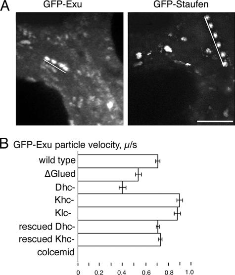
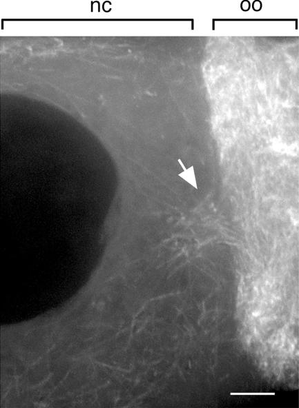
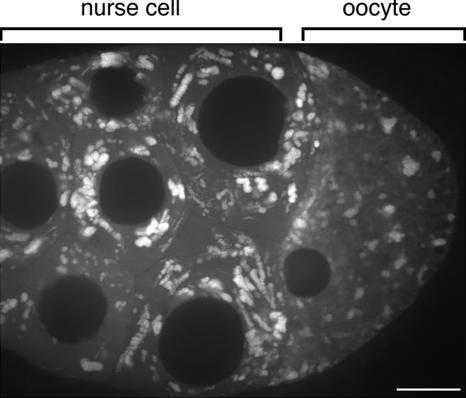
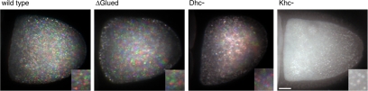
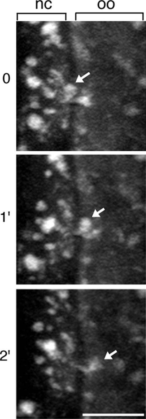
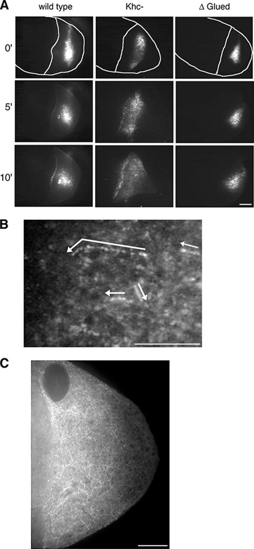
In Drosophila, the asymmetric localization of specific mRNAs to discrete regions within the developing oocyte determines the embryonic axes. The microtubule motors dynein and kinesin are required for the proper localization of the determinant ribonucleoprotein (RNP) complexes, but the mechanisms that account for RNP transport to and within the oocyte are not well understood. In this work, we focus on the transport of RNA complexes containing bicoid (bcd), an anterior determinant. We show in live egg chambers that, within the nurse cell compartment, dynein actively transports green fluorescent protein-tagged Exuperantia, a cofactor required for bcd RNP localization. Surprisingly, the loss of kinesin I activity elevates RNP motility in nurse cells, whereas disruption of dynein activity inhibits RNP transport. Once RNPs are transferred through the ring canal to the oocyte, they no longer display rapid, linear movements, but they are distributed by cytoplasmic streaming and gradually disassemble. By contrast, bcd mRNA injected into oocytes assembles de novo into RNP particles that exhibit rapid, dynein-dependent transport. We speculate that after delivery to the oocyte, RNP complexes may disassemble and be remodeled with appropriate accessory factors to ensure proper localization.


Figures

Figure 1.
Figure 1.
Quantification of GFP-RNP motility in nurse cells. (A) Time-lapse projections highlight linear movements of GFP-Exu (4 s) and GFP-Staufen (6 s) (Supplemental Movies S1 and S2). White lines indicate the path of a single particle within nurse cells. Bar, 5 μm. (B) Comparison of average velocities of GFP-Exu transport events in the nurse cells of wild-type, mutant, and colcemid-treated egg chambers. Disruption of dynein and kinesin I have opposite effects on the calculated velocities. Effects of the dynein and kinesin I mutations are fully rescued by their respective wild-type transgenes. On treatment with colcemid, GFP-Exu RNPs failed to undergo transport. Similar results were obtained for GFP-Staufen (Table 1). Dhc-, Dhc6-6/Dhc6-12; Khc-, Khc27; Klc-, klc8ex94. Error bars indicate SE of the mean.
Figure 2.
Figure 2.
Microtubule distribution at the ring canals leading into the oocyte. Within the nurse cells (nc), microtubules seem to be oriented randomly; however, near the ring canals (arrow), the number and organization of microtubules increases. Shown is a projection of eight 0.5-μm optical sections. oo, oocyte, Bar, 5 μm.
Figure 3.
Figure 3.
The compartmentalization of rapid cytoplasmic movements is illustrated by this 3-min time-lapse projection of GFP-Exu particles. Within the nurse cells, the RNPs exhibit robust linear transport. By contrast, in the oocyte, linear cytoplasmic movements of RNPs are not detectable over this timeframe (Supplemental Movie S3). The same phenomenon was observed for GFP-Staufen. Bar, 20 μm.
Figure 4.
Figure 4.
Cytoplasmic streaming does not require dynein. Three sequential images of autofluorescent yolk particles in stage 10b egg chambers were pseudocolored red, green, or blue with MetaMorph software and then superimposed. The resulting images reveal the presence or absence of movement as determined by the presence or absence of color. Moving particles display the red, green, blue color sequence, whereas the superimposition of images of nonmotile particles occurs as a single white particle. ΔGl and Dhc6-6/Dhc6-12 oocytes show a color pattern similar to wild-type, but Khc27 egg chambers show no movement in the oocyte (Supplemental Movie S4). Bar, 20 μm. Insets show 2.5× magnification.
Figure 5.
Figure 5.
RNP disassembly. After transport into the oocyte, GFP-Exu particles seem to decrease in size and/or disappear. Optical sections spanned sufficient depth to ascertain that the particle did not simply leave the focal plane. Arrow highlights a single particle undergoing this process as it enters the oocyte (Supplemental Movie S5). Bar, 10 μm, nc, nurse cell; oo, oocyte.
Figure 6.
Figure 6.
Fluorescently labeled bcd mRNA. (A) bcd RNA injected to the oocyte accumulates to the cortex in wild-type and Khc27 egg chambers. In contrast, this cortical accumulation is blocked in the UASp-ΔGl background. Still images from acquired time-lapse sequences at 0, 5, and 10 min after injection of bcd (Supplemental Movies S6–S8). Tracings of the egg chamber highlight the oocyte cortex. Bar, 20 μm. Throughout this figure, anterior is to the left, and posterior is to the right. (B) A time-lapse projection of 26 images reveals the tracks of bcd mRNA particles moving in a linear manner. Arrows indicate their direction of transport. Particles corresponding to the arrows are enhanced for clarity in this still image. Supplemental Movie S9. Bar, 10 μm. (C) Microtubule distribution within the oocyte as revealed with UAS-α-tubulin-GFP. Note the meshwork appearance of the microtubule array. A higher concentration of microtubules is present at the anterior cortex, compared with the posterior cortex. Bar, 10 μm.

References

    1. Berleth T., Burri M., Thoma G., Bopp D., Richstein S., Frigerio G., Noll M., Nusslein-Volhard C. The role of localization of bicoid RNA in organizing the anterior pattern of the Drosophila embryo. EMBO J. 1988;7:1749–1756. - PMC - PubMed
    1. Boylan K. L., Hays T. S. The gene for the intermediate chain subunit of cytoplasmic dynein is essential in Drosophila. Genetics. 2002;162:1211–1220. - PMC - PubMed
    1. Boylan K., Serr M., Hays T. A molecular genetic analysis of the interaction between the cytoplasmic dynein intermediate chain and the glued (dynactin) complex. Mol. Biol. Cell. 2000;11:3791–3803. - PMC - PubMed
    1. Brand A. H., Perrimon N. Targeted gene expression as a means of altering cell fates and generating dominant phenotypes. Development. 1993;118:401–415. - PubMed
    1. Brendza R. P., Serbus L. R., Duffy J. B., Saxton W. M. A function for kinesin I in the posterior transport of oskar mRNA and Staufen protein. Science. 2000;289:2120–2122. - PMC - PubMed

Publication types

MeSH terms

LinkOut - more resources