. 2007 Jun 15;581(Pt 3):1283-92.
doi: 10.1113/jphysiol.2007.129759. Epub 2007 Apr 12.

Effects of a R133W beta-tropomyosin mutation on regulation of muscle contraction in single human muscle fibres

Affiliations

Effects of a R133W beta-tropomyosin mutation on regulation of muscle contraction in single human muscle fibres

Julien Ochala et al. J Physiol. .

Abstract

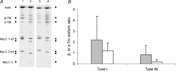
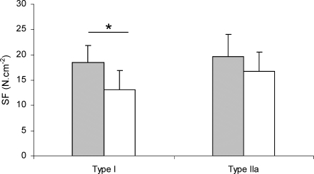
A novel R133W beta-tropomyosin (beta-Tm) mutation, associated with muscle weakness and distal limb deformities, has recently been identified in a woman and her daughter. The muscle weakness was not accompanied by progressive muscle wasting or histopathological abnormalities in tibialis anterior muscle biopsy specimens. The aim of the present study was to explore the mechanisms underlying the impaired muscle function in patients with the beta-Tm mutation. Maximum force normalized to fibre cross-sectional area (specific force, SF), maximum velocity of unloaded shortening (V0), apparent rate constant of force redevelopment (ktr) and force-pCa relationship were evaluated in single chemically skinned muscle fibres from the two patients carrying the beta-Tm mutation and from healthy control subjects. Significant differences in regulation of muscle contraction were observed in the type I fibres: a lower SF (P<0.05) and ktr (P<0.01), and a faster V0 (P<0.05). The force-pCa relationship did not differ between patient and control fibres, indicating an unaltered Ca2+ activation of contractile proteins. Collectively, these results indicate a slower cross-bridge attachment rate and a faster detachment rate caused by the R133W beta-Tm mutation. It is suggested that the R133W beta-Tm mutation induces alteration in myosin-actin kinetics causing a reduced number of myosin molecules in the strong actin-binding state, resulting in overall muscle weakness in the absence of muscle wasting.


Figures

Figure 1
Figure 1
α- and β-tropomyosin isoform expression A, electrophoretic separation of myosin heavy chain (MyHC), actin, and α- and β-tropomyosin (Tm) isoforms in single muscle fibres expressing type I (lanes 1 and 3) and IIa (lanes 2 and 4) MyHC isoforms from controls (lanes 1 and 2) and patients (lanes 3 and 4). B, β- to α-Tm isoform ratio in single muscle fibres from patients carrying the β-Tm mutation (open bar: type I fibres, n = 28; type IIa fibres, n = 8) and control subjects (hatched bar: type I fibres, n = 22; type IIa fibres, n = 8). Values are means ± s.d.
Figure 2
Figure 2
Force–pCa relationships Force–pCa curves of type I fibres from patients carrying the β-Tm mutation (•, n = 19) and control subjects (○, n = 20). All values are means.
Figure 3
Figure 3
Specific force SF, specific force. Comparison between fibres from patients carrying the β-Tm mutation (open bar: type I fibres, n = 51; type IIa fibres, n = 10) and those from control subjects (filled bar: type I fibres, n = 45; type IIa fibres, n = 29). Values are means ± s.d.*Statistically significant difference.
Figure 4
Figure 4
Apparent rate constant of force redevelopment (ktr) A, comparison between type I fibres from patients carrying the β-Tm mutation (open bar, n = 10) and control subjects (hatched bar, n = 10). Values are means ± s.d.*Statistically significant difference. B, typical experimental force signal recording from a type I fibre from a control subject (ktr 17.33 s−1). C, typical experimental force signal recording from a patient carrying the β-Tm mutation (ktr 7.70 s−1). The scale bar denotes 400 ms. A 20% release of original fibre length rapidly introduced at one end of the fibre reduces the proportion of available cross-bridges that are attached from approximately 80% in the isometric fibre to 20% (force drops to zero). Dissociation of the remaining cross-bridges is accomplished by rapidly re-extending overall muscle length to the initial value. Coincident with this re-stretch, force transiently increased because of positive straining of attached cross-bridges. These positively strained cross-bridges rapidly dissociated since the imposed length change is much greater than estimates of the working distance of a cross-bridge.
Figure 5
Figure 5
Maximum unloaded shortening velocity (V0) A, comparison between fibres from patients carrying the β-Tm mutation (open bar: type I fibres, n = 32; type IIa fibres, n = 8) and from control subjects (hatched bar: type I fibres, n = 25, type IIa fibres, n = 14). Values are means ± s.d.*Statistically significant difference. B, typical experimental force signal recording from a type I fibre from a control subject (12% slack, fibre length 1890 μm, V0 0.65 ML s−1 (muscle length per second)). C, typical experimental force signal recording from a patient carrying the β-Tm mutation (12% slack, fibre length 1800 μm, V0 1.27 ML s−1). The scale bar denotes 500 ms. When a release is introduced at one end of the fibre, force abruptly falls to zero, and the fibre is permitted to shorten under unloaded conditions.
Figure 6
Figure 6
The β-Tm mutation TnT binds Tm in an overall antiparallel manner; i.e. its C-terminal region is positioned near residue 190 of Tm, and the N-terminal tail of TnT extends towards the C terminus of Tm, overlapping the head to tail joint of Tm as well as 10–30 residues of the N terminus of the next molecule along the filament. The present β-TM mutation is located on residue 133.

Comment in

References

    1. Billeter R, Heizmann CW, Reist U, Howald H, Jenny E. α- and β-tropomyosin in typed single fibers of human skeletal muscle. FEBS Lett. 1981;132:133–136. - PubMed
    1. Bottinelli R, Coviello DA, Redwood CS, Pellegrino MA, Maron BJ, Spirito P, Watkins H, Reggiani C. A mutant tropomyosin that causes hypertrophic cardiomyopathy is expressed in vivo and associated with an increased calcium sensitivity. Circ Res. 1998;82:106–115. - PubMed
    1. Brandt PW, Diamond MS, Rutchik JS, Schachat FH. Co-operative interactions between troponin–tropomyosin units extend the length of the thin filament in skeletal muscle. J Mol Biol. 1987;195:885–896. - PubMed
    1. Brenner B. Effect of Ca2+ on cross-bridge turnover kinetics in skinned single rabbit psoas fibers: implications for regulation of muscle contraction. Proc Natl Acad Sci U S A. 1988;85:3265–3269. - PMC - PubMed
    1. Brenner B, Eisenberg E. Rate of force generation in muscle: correlation with actomyosin ATPase activity in solution. Proc Natl Acad Sci U S A. 1986;83:3542–3546. - PMC - PubMed

Publication types

MeSH terms

LinkOut - more resources