. 2007 Jul 1;582(Pt 1):95-111.
doi: 10.1113/jphysiol.2007.127894. Epub 2007 Apr 12.

Light triggers expression of philanthotoxin-insensitive Ca2+-permeable AMPA receptors in the developing rat retina

Affiliations

Light triggers expression of philanthotoxin-insensitive Ca2+-permeable AMPA receptors in the developing rat retina

Ingrid K Osswald et al. J Physiol. .

Abstract

Ca2+-permeable AMPA receptors (AMPARs) are expressed throughout the adult CNS but yet their role in development is poorly understood. In the developing retina, most investigations have focused on Ca2+ influx through NMDARs in promoting synapse maturation and not on AMPARs. However, NMDARs are absent from many retinal cells suggesting that other Ca2+-permeable glutamate receptors may be important to consider. Here we show that inhibitory horizontal and AII amacrine cells lack NMDARs but express Ca2+-permeable AMPARs. Before eye-opening, AMPARs were fully blocked by philanthotoxin (PhTX), a selective antagonist of Ca2+-permeable AMPARs. After eye-opening, however, a subpopulation of Ca2+-permeable AMPARs were unexpectedly PhTX resistant. Furthermore, Joro spider toxin (JSTX) and IEM-1460 also failed to antagonize, demonstrating that this novel pharmacology is shared by several AMPAR channel blockers. Interestingly, PhTX-insensitive AMPARs failed to express in retinae from dark-reared animals demonstrating that light entering the eye triggers their expression. Eye-opening coincides with the consolidation of inhibitory cell connections suggesting that the developmental switch to a Ca2+-permeable AMPAR with novel pharmacology may be critical to synapse maturation in the mammalian retina.


Figures

Figure 2
Figure 2. Horizontal and AII amacrine cells express synaptic AMPARs but not NMDARs
A, videomicrograph showing the position of the cell body close to the IPL (see arrowhead) and prominent dendrite characteristic of AII amacrine cells (see triangles) in acute retinal slice preparations. B, videomicrograph identifying the large triangular cell body of a typical horizontal cell (see arrowhead) and lateral dendritic processes (see triangles). C, whole-cell recording (Vh=−60 mV) from an AII cell showing spontaneous postsynaptic events (○) and inward current elicited by local application of 50 μm KA (cell no. 051208c3). Bath application of the non-NMDAR antagonist, CNQX (20 μm), blocked both spontaneous synaptic events and membrane current elicited by KA (•). D, whole-cell recording (Vh=−60 mV) from a horizontal cell showing that the spontaneous activity and holding current were insensitive to APV (40 μm), but blocked by GYKI 52466 (40 μm) (cell no. 060620c1). E, spontaneous events recorded from another AII amacrine cell (cell no. 060516c3, Vh=−60 mV) were insensitive to APV but blocked by GYKI 52466. F, spontaneous events recorded from the same horizontal cell as in D showing in more detail that synaptic activity was insensitive to APV but blocked by GYKI 52466.
Figure 6
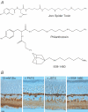
Figure 6. Retinal Ca2+-permeable AMPARs are insensitive to a number of channel blockers
A, extended structure of PhTX, JSTX and IEM-1460 reveals differences in their polyamine chain length as well as the chemical nature of their bulky headgroup. B, photomicrographs comparing the effect of 50 μm PhTX (slide no. 081206-8R), 10 μm JSTX (slide no. 081206-9R) and 100 μm IEM-1460 (slide no. 081206-7L) on the intensity of Co2+ labelling elicited by 10 mm Glu (slide no. 081206-9L) in the same P26 retina. Like PhTX, channel blockers JSTX and IEM-1460 also failed to block staining in the IPL as well as cell bodies of horizontal and AII amacrine cells.
Figure 7
Figure 7. Synaptic AMPARs expressed by AII cells are resistant to PhTX block
A, upper, synaptic activity (Vh=−70 mV) recorded from an AII amacrine cell (cell no. 050131c2) in control conditions. Lower, summary plot from 7 cells showing that the amplitude of miniature synaptic events was fitted well by the sum of three Gaussian functions (red line). Individual Gaussian fits are shown as continuous black lines. Similar results were observed in the absence of TTX (data not shown). B, upper, in the same cell, bath application of 50 μm PhTX did not fully block synaptic activity but instead reduced peak response amplitude. Lower, in this case, the summary plot of peak synaptic current amplitude was fitted well by the sum of two Gaussian functions (red line). Individual Gaussian fits are shown as continuous white lines. C, plot summarizing the effect of PhTX on the amplitude of spontaneous (n= 4) and miniature (n= 7) synaptic events from 11 cells. In all cases, analysis was performed only in cells where synaptic events were recorded in presence and absence of 50 μm PhTX. Data have been grouped on whether recordings were performed on postnatal retina of 3 weeks (n= 6) or more (n= 5). Filled (control) and open (+ 50 μm PhTX) symbols (triangle, circle and square) in each column represent the value of the peak amplitude obtained from individual Gaussian fits. Note PhTX failed to fully block the synaptic activity in all AII amacrine cell recordings.
Figure 10
Figure 10. AMPAR synaptic activity in AII amacrine cells is abolished by PhTX in dark-reared animals
A, synaptic activity (Vh=−70 mV) recorded from an AII amacrine cell (cell no. 061117c2) in a retina taken from a dark-reared animal. B, in the same cell, bath application of 50 μm PhTX fully blocked synaptic activity in contrast to the light-adapted retina. C, summary plot from 8 cells showing that the peak amplitude of miniature synaptic events was fitted well by the sum of three Gaussian functions (red line). Individual Gaussian fits are shown as continuous black lines. D, plot summarizing the effect of PhTX on the amplitude of spontaneous AMPAR synaptic events recorded from dark-reared retina (n= 4). Filled (control) symbols (triangle, circle and square) in each column represent the value of the peak amplitude obtained from individual Gaussian fits.
Figure 1
Figure 1. Ca2+-permeable AMPARs are expressed by inhibitory cells of the mammalian retina
Left panels, photomicrograph of Co2+ staining routinely observed with 10 mm l-Glu in a 3-month-old adult rat retina (slide no. 290306-1L). Staining was restricted to processes of the outer and inner plexiform layers as well as cell bodies of horizontal and AII amacrine cells. Co2+ staining elicited by 10 mm l-Glu was abolished by pretreatment with 40 μm GYKI 52466, an AMPAR selective antagonist. Middle upper panel, photomicrograph of calbindin immunofluorescence identifying the cell bodies and processes of horizontal cells. Middle panel, lower right and left, photomicrographs of visually identified AII amacrine cells filled with biocytin to reveal the location of the cell body in the INL and extension of dendritic processes. Right panels, Co2+ staining elicited by 10 mm l-Glu was unaffected by pretreatment with NMDA or the mGluR antagonists, APV (40 μm) (slide no. 070406-3L) and CPPG (300 μm) (slide no. 070406-9L), respectively. NMDA and group III mGluR agonists, NMDA (50 μm) (slide no. 070406-5R) and L-AP4 (100 μm) (slide no. 070406-9L), respectively, also failed to produce appreciable staining suggesting that inhibitory cells accumulate Co2+ due to the activation of AMPARs. Unless otherwise stated, scale bars represent 20 μm.
Figure 3
Figure 3. Expression of Ca2+-permeable AMPARs before eye-opening
Top left, outer and inner panels, cresyl violet staining of retina reveals an immature retina morphology at P1 (slide no. 090405-3L) with a prominent neuroblastic layer compared with the well-defined cell layers seen at P11 (slide no. 190405-5L). Middle left, outer and inner panels, Co2+ staining at P1 (slide no. 090405-7L) was blocked by 50 μm PhTX revealing the presence of Ca2+-permeable AMPARs at birth. Bottom left, outer and inner panels, Co2+ staining with 10 mm l-Glu in P4 (slide no. 120405-6L) and P7 (slide no. 150405-10L) retinae showing the migration and maturation of horizontal and amacrine cells. Middle and right panels, Co2+ staining elicited by l-Glu at P11 (slide no. 190405-2R) failed to be completely blocked by 50 μm PhTX suggesting that Ca2+-permeable AMPARs develop insensitivity to polyamine block a few days before eye-opening. Scale bars represent 10 μm in all cases.
Figure 4
Figure 4. Inhibitory cells express PhTX-insensitive Ca2+-permeable AMPARs after eye-opening
Left panels, after eye-opening horizontal and AII amacrine cells were strongly labelled by Co2+ at all developmental stages tested (P14–18, slide no. 220405-7R; P21–28, slide no. 250405-8R; P33–6 months: slide no. 260105-11L). Expression patterns in the INL are further refined during development with the transient expression of Ca2+-permeable AMPARs in another amacrine cell population(s) that may be A17 cells. Note there is a greater intensity of staining in sublamina b of the IPL (see arrowhead). Right panels, PhTX failed to fully block Co2+ staining after eye-opening at all developmental stages. Horizontal cell dendrites in particular were completely insensitive to block by PhTX. For AII cells, Co2+ staining of cell bodies was initially PhTX insensitive (P14–18, slide no. 220405-7R; P21–28, slide no. 250405-8R, respectively). However, at later stages, the staining of cell bodies was abolished by PhTX. In contrast, staining of the IPL was resistant to PhTX block at all stages.
Figure 5
Figure 5. AII amacrine cell dendrites express two AMPAR populations
A, left, photomicrograph of an AII amacrine cell filled with biocytin reveals the extent of its dendritic arborization in the IPL. Right, schematic diagram showing the possible spatial arrangement of different AMPARs on dendrites of individual AII cells. B, left, photomicrographs comparing the effect of 50 μm PhTX (slide no. 260105-11R) on the staining elicited by 10 mm Glu (slide no. 260105-11R) in the IPL of a P36 retina. The dotted line demarcates between sublaminae a and b and the scale bar represents 20 μm. Right, images from bright-field microscope were obtained in black and white format and measurements of the relative optical density were determined for the OPL and sublaminae a and b of the IPL using an MCID Elite Image Analysis system (see Methods). A total number of eight images were obtained per treatment which was repeated for retinae taken from 8 adult rats. The data between different treatments were compared using a paired t test and expressed as the mean ±s.e.m.
Figure 8
Figure 8. PhTX slows decay kinetics of synaptic AMPAR events
A, averaged mEPSCs from the same cell show differences in decay kinetics before and after application of PhTX (cell no. 050318c1). B–D, summary plots showing that the decay kinetics of synaptic events before (B) and after (C) PhTX application (n= 7) are different. D, the kinetic properties of events less than –20 pA are indistinguishable from the entire event population suggesting that PhTX slows decay kinetics of synaptic AMPARs.
Figure 9
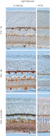
Figure 9. Inhibitory cells fail to express PhTX-insensitive Ca2+-permeable AMPARs in dark-reared animals
Left panels, Co2+ staining pattern of horizontal and AII amacrine cells from dark-reared animals was similar to the light-adapted retina (P14–18, slide no. 010206-8R; P21–28, slide no. 050605-10L; P33–6 months, slide no. 210206-8L). Right panels, in contrast, however, 50 μm PhTX completely eliminated staining of AII cells and the IPL and significantly reduced Co2+ labelling of horizontal cells (P14–18, slide no. 010206-6L; P21–28, slide no. 050605-7L; P33–6 months, slide no. 210206-7R).
Figure 11
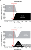
Figure 11. Expression of Ca2+-permeable AMPARs is triggered by light entering the eye
Schematic diagrams intended to provide a qualitative description of the expression profile of Ca2+-permeable AMPARs in AII and horizontal cells. The transient expression of Ca2+-permeable AMPARs in other cells of the INL and GCL has not been included since it was not studied in detail. A, in horizontal cells, Ca2+-permeable AMPARs sensitive to PhTX were replaced by AMPARs with little or no sensitivity to external polyamines after eye-opening. B, for AII amacrine cells, the expression profile is more complex. Prior to eye-opening, the cell bodies and dendrites of AII cells express PhTX-sensitive AMPARs. Like horizontal cells, AMPARs that are insensitive to PhTX are detected just before eye-opening (see arrows) and continue to be expressed on cell bodies and dendrites of AII cells in the first 3–4 weeks postnatal. However as development proceeds, the expression of PhTX-insensitive AMPARs is restricted to the IPL (hatched bar).

Comment in

Similar articles

Cited by

References

    1. Aizenman E, Karschin A, Lipton SA. Two pharmacological classes of quisqualate-induced electrical responses in rat retinal ganglion cells in vitro. Eur J Pharmacol. 1989;174:9–22. - PubMed
    1. Bähring R, Mayer ML. An analysis of philanthotoxin block for recombinant rat GluR6(Q) glutamate receptor channels. J Physiol. 1998;509:635–650. - PMC - PubMed
    1. Bellone C, Luscher C. Cocaine triggered AMPA receptor redistribution is reversed in vivo by mGluR-dependent long-term depression. Nat Neurosci. 2006;9:636–641. - PubMed
    1. Blaschke M, Keller BU, Rivosecchi R, Hollmann M, Heinemann S, Konnerth A. A single amino acid determines the subunit-specific spider toxin block of alpha-amino-3-hydroxy-5-methylisoxazole-4-propionate/kainate receptor channels. Proc Natl Acad Sci U S A. 1993;90:6528–6532. - PMC - PubMed
    1. Bowie D, Bähring R, Mayer ML. Block of kainate and AMPA receptors by polyamines and arthropod toxins. In: Jonas P, Monyer H, editors. Handbook of Experimental Pharmacology; Ionotropic Glutamate Receptors in the CNS. Berlin: Springer-Verlag; 1999. pp. 251–373.

Publication types

MeSH terms

LinkOut - more resources