. 2007 May 2;26(9):2350-60.
doi: 10.1038/sj.emboj.7601684. Epub 2007 Apr 12.

XTsh3 is an essential enhancing factor of canonical Wnt signaling in Xenopus axial determination

Affiliations

XTsh3 is an essential enhancing factor of canonical Wnt signaling in Xenopus axial determination

Takayuki Onai et al. EMBO J. .

Abstract

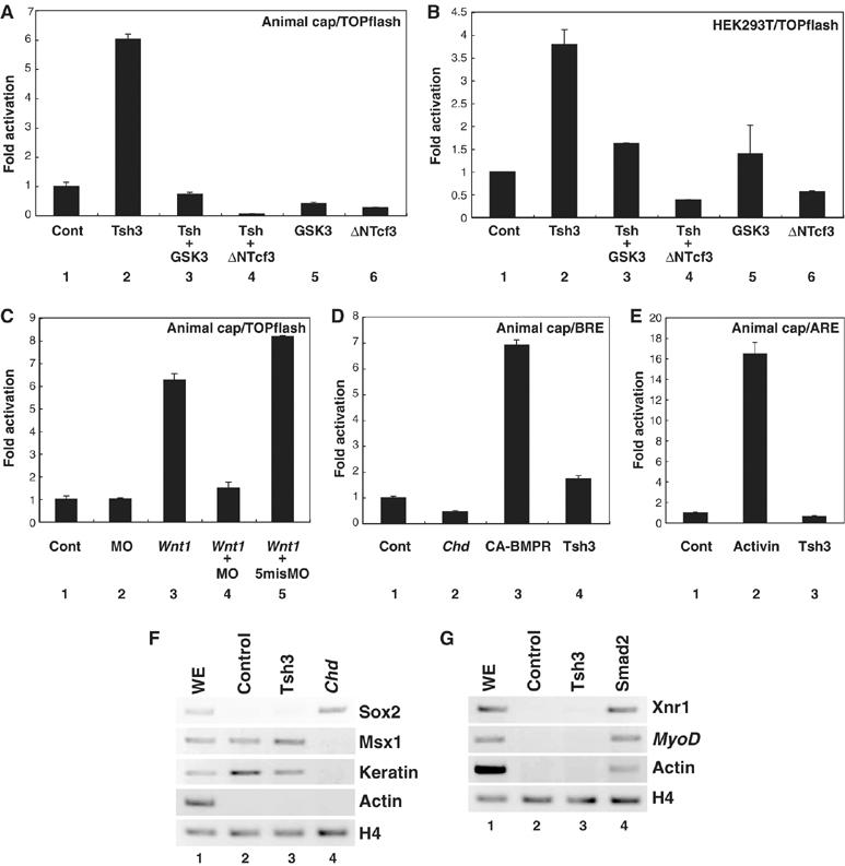
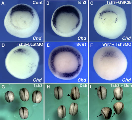
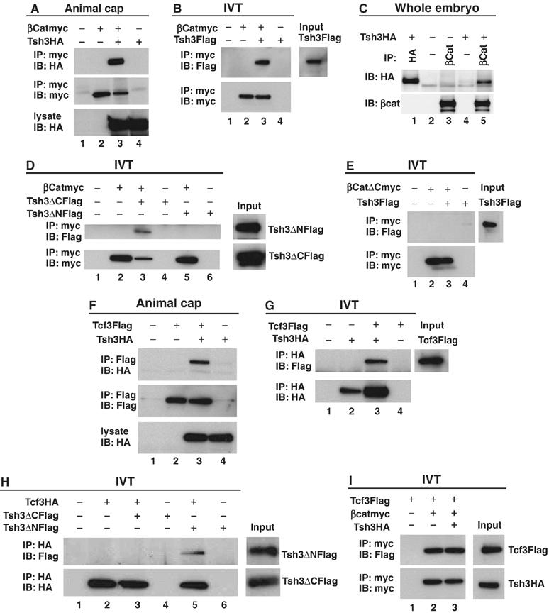
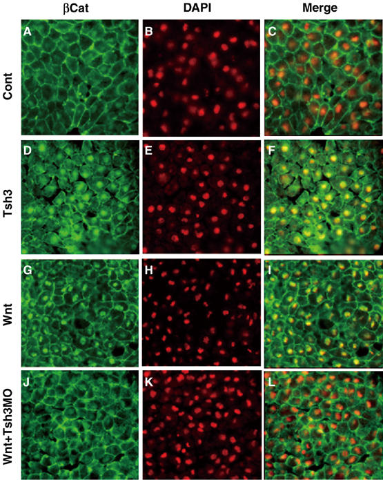
In Xenopus, an asymmetric distribution of Wnt activity that follows cortical rotation in the fertilized egg leads to the dorsal-ventral (DV) axis establishment. However, how a clear DV polarity develops from the initial difference in Wnt activity still remains elusive. We report here that the Teashirt-class Zn-finger factor XTsh3 plays an essential role in dorsal determination by enhancing canonical Wnt signaling. Knockdown of the XTsh3 function causes ventralization in the Xenopus embryo. Both in vivo and in vitro studies show that XTsh3 substantially enhances Wnt signaling activity in a beta-catenin-dependent manner. XTsh3 cooperatively promotes the formation of a secondary axis on the ventral side when combined with weak Wnt activity, whereas XTsh3 alone has little axis-inducing ability. Furthermore, Wnt1 requires XTsh3 for its dorsalizing activity in vivo. Immunostaining and protein analyses indicate that XTsh3 is a nuclear protein that physically associates with beta-catenin and efficiently increases the level of beta-catenin in the nucleus. We discuss the role of XTsh3 as an essential amplifying factor of canonical Wnt signaling in embryonic dorsal determination.


Figures

Figure 1
Figure 1
Deduced protein structure and expression profile of XTsh3. (A) Deduced domain structure of the XTsh3 protein. Blue, Zn-finger motif; gray, Hox domain; and black, basic motif. (B–I) Whole-mount in situ hybridization analysis of XTsh3 expression. (B, C) Maternal expression in the animal region at the eight-cell stage. Animal (B) and lateral (C) views. (D) Dorsally dominant expression at stage 11. Vegetal view. Arrowhead, dorsal lip. (E, F) stage 12.5, dorsal (E) and ventral (F) views. Arrowhead, closing blastopore. Section analysis also showed strong XTsh3 expression in the dorsal mesoderm and ectoderm (not shown). (G) Stage 18, dorsal view. (H, I) Tailbud (stage 24) and early larval (stage 31) stages, lateral views. Arrowhead, anterior expression border at the hindbrain. Strong XTsh3 expression gradually became limited to the caudal CNS.
Figure 2
Figure 2
XTsh3 is essential for dorsal axial development in Xenopus. (A–C) Ventralization by XTsh3-MO. Injection of XTsh3-MO (MO-A, 20 ng/cell; B) into two dorsal blastomeres of four-cell embryos depleted the dorsal axes, while such ventralization was not seen with ventral XTsh3-MO injection (C). (D–K) Effects of radial XTsh3-MO injection on DV marker gene expression (D, E, Chd; F, G, Gsc; H, I, Vent2; J, K, Myf5) at the gastrula stage shown by in situ hybridization. Cont, control injection. Open arrowheads, dorsal borders of Vent2 expression. (L, M) Chd expression suppressed with XTsh3-MO (E) was rescued by coinjection of mTsh1 (L). (M) mTsh1 injection alone laterally expanded Chd expression (D, M). (N) RT–PCR of DMZ explants (stage 13 equivalent). Lane 1, whole embryo; lane 2, control DMZ; and lane 3, XTsh3-MO-injected DMZ.
Figure 3
Figure 3
Dorsalizing activity of XTsh3. Whole-mount in situ hybridization analysis of DV marker gene expression (A, B, Chd; C, D, Gsc; E, F, Szl; G, H, Vent1) in control (A, C, E, G) and XTsh3-injected (400 pg/cell, four-cell stage; B, D, F, H) gastrula embryos. Vegetal view with the dorsal side up. The expression of the ventral markers was reduced and narrowed (indicated by arrowheads) by XTsh3 injection.
Figure 4
Figure 4
XTsh3 is required for efficeint canonical Wnt signaling. (A–C) Topflash-luciferase assay using animal caps (A, C) and 293T cells (B). (A, B) RNAs (A) or expression plasmids (B) used: lane 1, control; lane 2, XTsh3; lane 3, XTsh3 and GSK3β; lane 4, XTsh3 and dnTcf3, lane 5, and GSK3β; lane 6, dnTcf3. (C) RNAs used: lane 1, control; lane 2, XTsh3-MO; lane 3, Wnt1; lane 4, Wnt1 and XTsh3-MO, and lane 5, Wnt1 and control MO. (D) BRE (BMP response element)-luciferase assay using animal caps. RNAs used: lane 1, control; lane 2, Chd; lane 3, CA-BMPR (constitutive active); and lane 4, XTsh3. (E) ARE (Activin response element) luciferase assay using animal caps. Lane 1, control; lane 2, Activin treatment; and lane 3, XTsh3. (F) RT–PCR analysis of neural and non-neural ectodermal marker genes in RNA-injected animal caps (stage 12 equivalent). Unlike Chd injection (lane 4), XTsh3 injection (lane 3) did not induce strong expression of the neural marker Sox2 or suppress the non-neural genes Msx1 and Keratin. (G) RT–PCR analysis of mesodermal marker genes in injected animal caps (stage 12 equivalent). Unlike Smad2 injection (lane 4; mimicking Activin signaling), XTsh3 injection (lane 3) did not induce strong expression of Xnr1, MyoD or cardiac actin.
Figure 5
Figure 5
XTsh3 enhances and requires Wnt/β-catenin signaling in dorsal determination. (A–F) Effects on Chd expression at the gastrula stages. (B–D) Expansion of Chd expression by radial XTsh3 injection (B) was reversed by coinjecting GSK3β RNA (100 pg/cell; C) and β-catenin-MO (20 ng/cell; D). (E and F) Strong induction of Chd by radial Wnt1 injection (25 pg/cell; E) was inhibited by coinjecting XTsh3-MO (20 ng/cell; F). (G–I) Double axis formation by Dsh injection into a ventral-vegetal blastomere at the eight-cell stage (analyzed at the late neurula stage). (G) XTsh3 (100 pg/cell), (H) Dsh (100 pg/cell), and (I) Dsh and XTsh3. Arrows, secondary axes and arrowheads, weak axes.
Figure 6
Figure 6
Protein interaction of XTsh3 with β-catenin. (A–D) Immunoprecipitation assay of XTsh3 and β-catenin. (A) Immunoprecipitation from animal cap extracts injected with XTsh3-HA (lanes 3 and 4) and/or β-catenin-myc (lanes 2 and 3). (B) Immunoprecipitation with in vitro translation products of XTsh3-Flag (lanes 3 and 4) and/or β-catenin-myc (lanes 2 and 3). (C) Immunoprecipitation of XTsh3 and endogenous β-catenin from the whole-embryo extract. Anti-β-catenin antibody (which binds to endogenous β-catenin) co-precipitated XTsh3-HA (lane 5; negative control in lane 3) while the control antibody (that does not bind to β-catenin) did not (lane 4). (D) Co-precipitation of in vitro translation products was seen with β-catenin-myc and XTsh3-ΔC-Flag (lane 3) but not with β-catenin-myc and XTsh3-ΔN-Flag (lane 5). (E) Co-precipitation of in vitro translation products was not seen with β-catenin-ΔC-myc and XTsh3-Flag (lane 3). (F–H) Immunoprecipitation assay of XTsh3 and Tcf3. (F) Immunoprecipitation from animal cap extracts injected with XTsh3-HA (lanes 3 and 4) and/or Tcf3-Flag (lanes 2 and 3). (G) Immunoprecipitation with in vitro translation products of XTsh3-HA (lanes 2 and 3) and/or Tcf3-Flag (lanes 3 and 4). (H) Co-precipitation of in vitro translation products was seen with Tcf3-HA and XTsh3-ΔN-Flag (lane 5) but not with Tcf3-HA and XTsh3-ΔC-Flag (lane 3). (I) Co-precipitation of Tcf3 and β-catenin was not affected by the presence of XTsh3 (lane 3).
Figure 7
Figure 7
XTsh3 is essential for the nuclear localization of β-catenin induced by Wnt. Immunostaining study of the nuclear accumulation of β-catenin protein in animal cap cells. Animal caps (excised at stage 9 and fixed at stage 11) were prepared from embryos injected with control (A–C), XTsh3-HA (D–F), Wnt1 (G–I), Wnt1 and XTsh3-MO (J–L). (A, D, G, J) Fluorescent immunostaining of endogenous β-catenin. (B, E, H, K) Nuclear DAPI staining. (C, F, I, L) Merged pictures of the β-catenin and DAPI signals.

Similar articles

Cited by

References

    1. Abe K, Takeichi M (2007) NMDA-receptor activation induces calpain-mediated beta-catenin cleavages for triggering gene expression. Neuron 53: 387–397 - PubMed
    1. Andrew JD, Horner MA, Petitt MG, Smolik SM, Scott MP (1994) Setting limits on homeotic gene function: restrint of Sex combs reduced activity by teashirt and other homeotic genes. EMBO J 13: 1132–1144 - PMC - PubMed
    1. Bessa J, Casares F (2005) Restricted teashirt expression confers eye-specific responsiveness to Dpp and Wg signals during eye specification in Drosophila. Development 132: 5011–5020 - PubMed
    1. Bessa J, Gebelein B, Pichaud F, Casares F, Mann RS (2002) Combinatorial control of Drosophila eye development by Eyeless, Homothorax, and Teashirt. Gene Dev 16: 2415–2427 - PMC - PubMed
    1. Brannon M, Gomperts M, Sumoy L, Moon RT, Kimelman D (1997) A beta-catenin/XTcf-3 complex binds to the siamois promoter to regulate dorsal axis specification in Xenopus. Genes Dev 11: 2359–2370 - PMC - PubMed

Publication types

LinkOut - more resources