. 2007 Apr;20(2):125-35.
doi: 10.1080/08998280.2007.11928267.

Thoracic outlet syndrome: a 50-year experience at Baylor University Medical Center

Affiliations

Thoracic outlet syndrome: a 50-year experience at Baylor University Medical Center

Harold C Urschel et al. Proc (Bayl Univ Med Cent). 2007 Apr.

Abstract

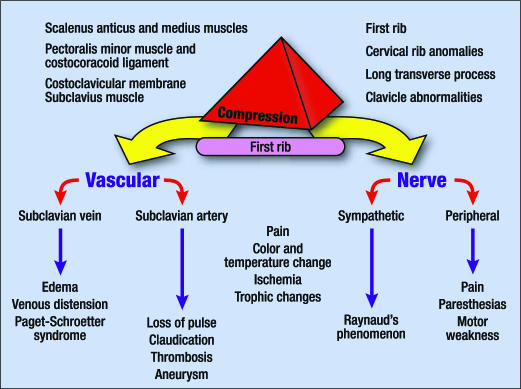
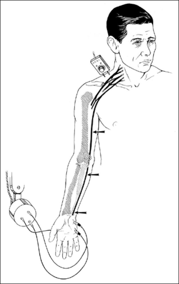
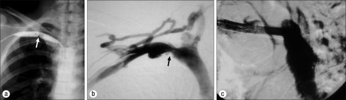
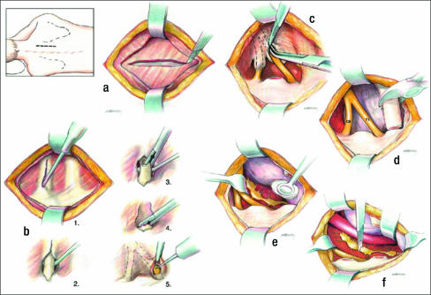
During the past 5 decades, the recognition and management of thoracic outlet syndrome (TOS) have evolved. This article elucidates these changes and improvements in the diagnosis and management of TOS at Baylor University Medical Center. The most remarkable change over the past 50 years is the use of nerve conduction velocity to diagnose and monitor patients with nerve compression. Recognition that procedures such as breast implantation and median sternotomy may produce TOS has been revealing. Prompt thrombolysis followed by surgical venous decompression for Paget-Schroetter syndrome has markedly improved results compared with the conservative anticoagulation approach; thrombolysis and prompt first rib resection is the optimal treatment for most patients with Paget-Schroetter syndrome. Complete first rib extirpation at the initial procedure markedly reduces the incidence of recurrent neurologic symptoms or the need for a second procedure. Chest pain or pseudoangina can be caused by TOS. Dorsal sympathectomy is helpful for patients with sympathetic maintained pain syndrome or causalgia and patients with recurrent TOS symptoms who need a second procedure.


Figures

Figure 1
Figure 1
Compression factors in the thoracic outlet with the signs and symptoms produced.
Figure 2
Figure 2
Neurovascular structures traversing the thoracic outlet. Netter illustration used with permission of Elsevier Inc. All rights reserved.
Figure 3
Figure 3
Cervical ribs and related anomalies. Netter illustration used with permission of Elsevier Inc. All rights reserved.
Figure 4
Figure 4
Nerve conduction velocity measurement technique. There is a “blip” on the oscilloscope when the electric current hits the electrode.
Figure 5
Figure 5
Transaxillary first rib resection. (a) Division of the scalenus anticus muscle. (b) Division of the first rib and anterior resection. (c) Posterior resection of the first rib. (d) Resection of head and neck of rib. (e) Identification of the dorsal sympathetic chain. (f) Division through the lower stellate ganglion above T1 and below T3 ganglia. V indicates subclavian vein; SA, scalenus anticus muscle; A, subclavian artery; BP, brachial plexus; SM, sternocleido-mastoid muscle.
Figure 6
Figure 6
Urschel first rib rongeur.
Figure 7
Figure 7
Supraclavicular approach. (a) Supraclavicular incision. (b) Division of the scalenus anticus muscle. (c) Supraclavicular retraction of the neurovascular structures. (d–f) Resection of first rib. (g) Supraclavicular dorsal sympathectomy. BP indicates brachial plexus; A, subclavian artery; SA, scalenus anticus muscle; P, phrenic nerve.
Figure 8
Figure 8
Examples of arterial compression resulting from thoracic outlet compression. (a) Poststenotic dilatation of the axillary-subclavian artery. (b) Sacular aneurysm of the axillary-subclavian artery. (c) Total occlusion of the axillary subclavian artery.
Figure 9
Figure 9
Paget-Schroetter syndrome. (a) Occlusion of the axillary-subclavian vein. (b) Swollen right hand.
Figure 10
Figure 10
Inappropriate treatment of Paget-Schroetter syndrome. (a) Pericutaneous balloon dilatation. (b) Venous recoil after balloon deflation. (c) Deleterious stent insertion.
Figure 11
Figure 11
Posterior reoperation for recurrent thoracic outlet syndrome. (a) Muscle-splitting incision. (b) Removal of in older patients with markedly rib remnant and neurolysis of C8, T1 nerve roots. (c–e) Neurolysis of nerve roots and posterior dorsal sympathectomy.

References

    1. Peet RM, Hendriksen JD, Anderson TP, Martin GM. Thoracic outlet syndrome: evaluation of the therapeutic exercise program. Proc Mayo Clin. 1956;31:281–287. - PubMed
    1. Borchardt M. Symptomatologie und Therapie der Halsrippen. Berl Klin Wochenschr. 1901;38:1265.
    1. Keen WW. The symptomatology, diagnosis and surgical treatment of cervical ribs. Am J Med Sci. 1907;133:173–218.
    1. Adson AW, Coffey JR. Cervical rib: a method of anterior approach for relief of symptoms by division of the scalenus anticus. Ann Surg. 1927;85:839–857. - PMC - PubMed
    1. Coote H. Pressure on the axillary vessels and nerves by an exostosis from a cervical rib; interference with the circulation of the arm; removal of the rib and exostosis, recovery. Med Times Gaz. 1861;2:108.

LinkOut - more resources