

Acute esophageal necrosis and low-flow state
- PMID: 17431514
- PMCID: PMC2657700
- DOI: 10.1155/2007/920716
Acute esophageal necrosis and low-flow state
Abstract
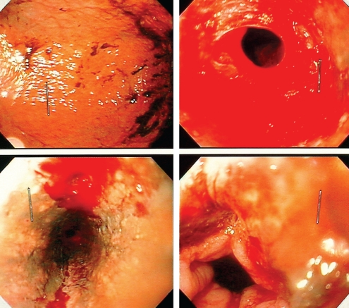
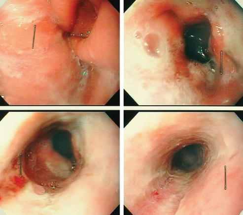
Acute esophageal necrosis (AEN), also called black esophagus, is quite exceptional. Endoscopic findings show circumferential black discolouration of the esophagus with or without exudates. The etiology of AEN is presently unknown and is assumed to be multifactorial. Distal esophageal involvement with proximal extension ending sharply at the gastroesophageal junction is the most common presentation. The present case report describes the clinical and endoscopic evolution of black esophagus observed in a patient with significant peripheral vascular disease, who was presented to the intensive care unit at the Hopital Saint-Francois d'Assise (Quebec City, Quebec). Through an extensive review of the literature, common underlying clinical conditions of patients diagnosed with AEN have been identified.
La nécrose œsophagienne aiguë (NOA), aussi appelée « œsophage noir », est un événement rare. Les examens endoscopiques révèlent une coloration noire, circonférentielle de l’œsophage, avec ou sans exsudat. On ne connaît pas l’étiologie de la NOA et on croit qu’elle est plurifactorielle. La maladie consiste le plus souvent en une atteinte distale de l’œsophage avec une extension proximale se terminant abruptement à la jonction oeso-gastrique. Le présent rapport décrit l’évolution d’un cas d’œsophage noir chez un patient atteint d’une grave maladie vasculaire périphérique admis aux soins intensifs de l’hôpital Saint-François d’Assise (Québec, Québec). Après un examen approfondi de la littérature, il a été possible de recenser les pathologies cliniques sous-jacentes souvent rencontrées chez les patients victimes de NOA.
Figures
Similar articles
-
Black esophagus: acute esophageal necrosis syndrome.World J Gastroenterol. 2010 Jul 14;16(26):3219-25. doi: 10.3748/wjg.v16.i26.3219. World J Gastroenterol. 2010. PMID: 20614476 Free PMC article. Review.
-
Black esophagus: new insights and multicenter international experience in 2014.Dig Dis Sci. 2015 Feb;60(2):444-53. doi: 10.1007/s10620-014-3382-1. Epub 2014 Oct 9. Dig Dis Sci. 2015. PMID: 25297468
-
[Acute esophageal necrosis: an unusual entity].Medicina (B Aires). 2010;70(6):524-6. Medicina (B Aires). 2010. PMID: 21163740 Spanish.
-
Black esophagus: a case series and literature review of acute esophageal necrosis.Scand J Gastroenterol. 2018 Oct-Nov;53(10-11):1421-1424. doi: 10.1080/00365521.2018.1513064. Epub 2018 Oct 24. Scand J Gastroenterol. 2018. PMID: 30353761 Review.
-
[Acute esophageal necrosis. An underdiagnosed disease].Rev Esp Enferm Dig. 2008 Nov;100(11):701-5. doi: 10.4321/s1130-01082008001100006. Rev Esp Enferm Dig. 2008. PMID: 19159174 Spanish.
Cited by
-
Acute Esophageal Necrosis: An Update.N Am J Med Sci. 2016 Jul;8(7):320-2. doi: 10.4103/1947-2714.187159. N Am J Med Sci. 2016. PMID: 27583242 Free PMC article.
-
A case of unusual presentation of acute esophageal necrosis with pneumonia.Int J Health Sci (Qassim). 2020 Nov-Dec;14(6):66-68. Int J Health Sci (Qassim). 2020. PMID: 33192233 Free PMC article.
-
Gurvits Syndrome: Black Esophagus in the Postoperative Setting.Cureus. 2022 Jan 14;14(1):e21240. doi: 10.7759/cureus.21240. eCollection 2022 Jan. Cureus. 2022. PMID: 35174035 Free PMC article.
-
Diagnosis and management of acute esophageal necrosis.Ann Gastroenterol. 2019 Nov-Dec;32(6):529-540. doi: 10.20524/aog.2019.0418. Epub 2019 Sep 26. Ann Gastroenterol. 2019. PMID: 31700229 Free PMC article. Review.
-
Black esophagus: acute esophageal necrosis syndrome.World J Gastroenterol. 2010 Jul 14;16(26):3219-25. doi: 10.3748/wjg.v16.i26.3219. World J Gastroenterol. 2010. PMID: 20614476 Free PMC article. Review.
References
-
- Carneiro M, Lescano M, Romanello L, et al. Acute esophageal necrosis. Dig Endosc. 2005;17:89–92.
-
- Soussan EB, Savoye G, Hochain P, et al. Acute esophageal necrosis: A 1-year prospective study. Gastrointest Endosc. 2002;56:213–7. - PubMed
-
- Goldenberg SP, Wain SL, Marignani P. Acute necrotizing esophagitis. Gastroenterology. 1990;98:493–6. - PubMed
-
- Moreto M, Ojembarrena E, Zaballa M, Tanago JG, Ibanez S. Idiopathic acute esophageal necrosis: Not necessarily a terminal event. Endoscopy. 1993;25:534–8. - PubMed
-
- Lacy BE, Toor A, Bensen SP, Rothstein RI, Maheshwari Y. Acute esophageal necrosis: Report of two cases and a review of the literature. Gastrointest Endosc. 1999;49:527–32. - PubMed
Publication types
MeSH terms
LinkOut - more resources
Full Text Sources
Miscellaneous