. 2007 Oct;56(10):1625-36.
doi: 10.1007/s00262-007-0309-0. Epub 2007 Apr 13.

Interleukin-4 impairs granzyme-mediated cytotoxicity of Simian virus 40 large tumor antigen-specific CTL in BALB/c mice

Affiliations

Interleukin-4 impairs granzyme-mediated cytotoxicity of Simian virus 40 large tumor antigen-specific CTL in BALB/c mice

Nikola Baschuk et al. Cancer Immunol Immunother. 2007 Oct.

Abstract

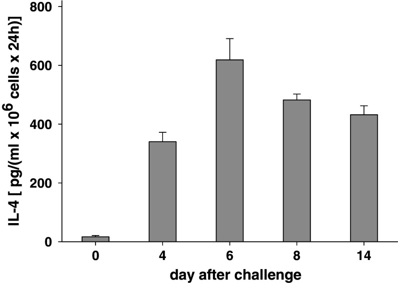
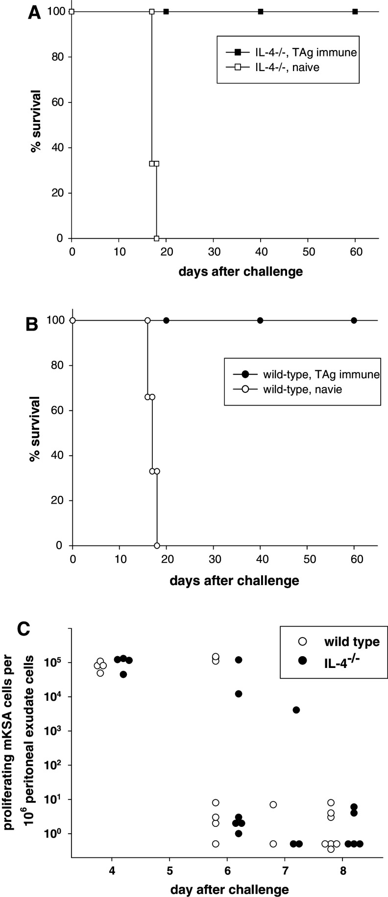
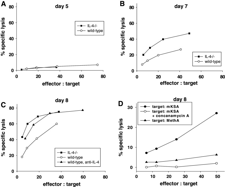
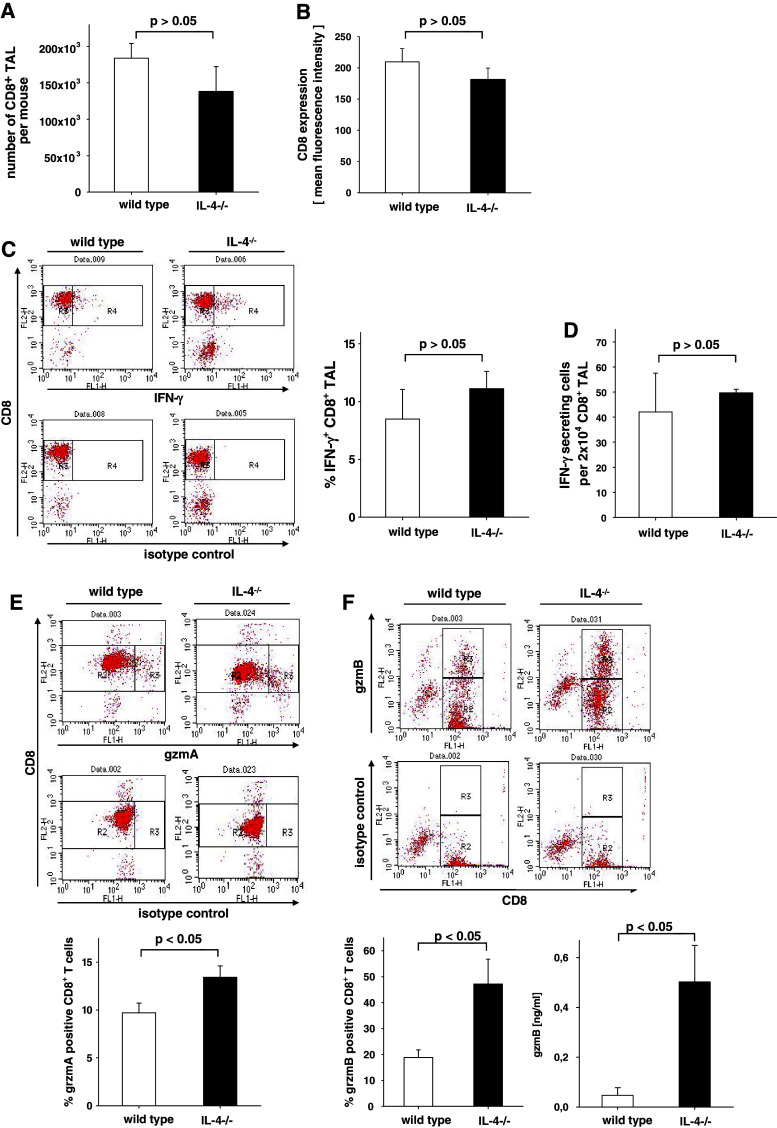
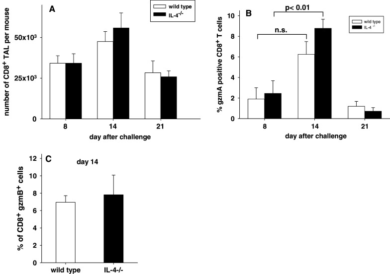
In this report we analyzed the impact of interleukin-4 (IL-4) on tumor-associated simian virus 40 (SV40) large T-antigen (TAg)-specific CD8+ cytotoxic T cells during rejection of syngeneic SV40 transformed mKSA tumor cells in BALB/c mice. Strikingly, challenge of naïve mice with low doses of mKSA tumor cells revealed a CD8+ T cell-dependent prolonged survival time of naïve IL-4-/- mice. In mice immunized with SV40 TAg we observed in IL-4-/- mice, or in wild type mice treated with neutralizing anti-IL-4 monoclonal antibody, a strongly enhanced TAg-specific cytotoxicity of tumor associated CD8+ T cells. The enhanced cytotoxicity in IL-4-/- mice was accompanied by a significant increase in the fraction of CD8+ tumor associated T-cells expressing the cytotoxic effector molecules granzyme A and B and in granzyme B-specific enzymatic activity. The data suggest that endogenous IL-4 can suppress the generation of CD8+ CTL expressing cytotoxic effector molecules especially when the antigen induces only a very weak CTL response.


Figures

Fig. 1
Fig. 1
Primary CD4+ TAL from TAg-immunized BALB/c mice secrete IL-4 after challenge with mKSA. BALB/c mice were immunized on days 14 and 7 by i.p. injection of 10 μg TAg and challenged i.p. on day 0 with 106 mKSA cells. CD4+ T cells were immunomagnetically enriched from peritoneal exudate cells collected prior to challenge (day 0; cells pooled from 15 mice) or on days 4, 6, 8, or 14 after challenge (pooled from five mice per day). The CD4+ TAL were kept at a densitiy of 1 × 106 cells/ml in complete RPMI medium without any further stimuli for 24 h. The contents of IL-4 in the cell-free supernatants of these cultures were determined by ELISA. Shown are the means and standard error of the mean of three independent experiments
Fig. 2
Fig. 2
TAg-immune IL-4−/− BALB/c mice are fully protected against challenge with 106 viable mKSA tumor cells. Groups of IL-4−/− and wt BALB/c mice were immunized on days −14 and −7 by i.p. injection of 10 μg TAg and challenged i.p. with 106 mKSA cells on day 0. Survival of (A) IL-4−/− and (B) wt mice (immune n = 5, non-immune n = 3) was monitored daily up to 65 days after challenge. (C) On the indicated days after challenge, peritoneal exudate cells harvested by peritoneal lavage were plated in ten-fold serial dilutions on 6-well plates. After cultivation for 8 days the cells were fixed and stained with crystal violett and clonal colonies of mKSA cells were counted. Shown are cumulative data from three independent experiments with each symbol representing an individual mouse
Fig. 3
Fig. 3
Tumor growth following s.c. inoculation and survival of mice after i.p. injection of mKSA cells into naive IL-4−/− and wild-type mice. a Naïve IL-4−/− (closed circles, n = 11) or wild-type mice (open circles, n = 12) were inoculated with 102 viable mKSA cells s.c. in the back of the neck. At the indicated days after challenge the diameters of the tumors were determined in three dimensions with calipers and subsequently the tumor volume was calculated. b Groups of naive IL-4−/− (closed symbols, n = 14) or wild-type mice (open symbols, n = 11) were i.p. inoculated with 103 viable mKSA cells. The mice were monitored for the development of tumors and death daily. According to Kaplan–Meier analysis the mean survival time and the standard error were for the IL-4−/− group 40.14 ± 4.38 days, and for the wild type 25.45 ± 2.23 days with a statistical significance of P < 0.01 in log-rank test. c Groups of naive IL-4−/− (n = 8) and wt (n = 10) mice were i.v. treated with 500 μg of either CD8-specific mAb or of control IgG 1 day prior to i.p. challenge with 103 mKSA cells. Treatment with the antibodies was repeated 8 days after challenge. The mice were monitored for the development of tumors and death daily. According to Kaplan–Meier analysis the mean survival time in days +/− standard error and the statistical significance in the log-rank test were as follows: IL-4−/−: anti-CD8 treated 25.88 ± 0.79 versus control IgG 51.13 ± 7.57, P < 0.01; wt: anti-CD8 treated 25.80 ± 1.29 versus control IgG 36.90 +/− 6.08, p > 0.05 in log-rank test
Fig. 4
Fig. 4
Primary cytotoxicity of CD8+ TAL from TAg-immunized IL-4−/− or wild-type mice at different times after challenge. IL-4−/− or wild-type mice were immunized on days 14 and 7 by i.p. injection of 10 μg TAg and challenged i.p. with 106 mKSA cells on day 0. CD8+ TAL were immunomagnetically enriched from the pooled peritoneal exudate cells of groups of five mice on day 5 (a), day 7 (b), or day 8 (c) after challenge. On day 8 CD8+ TAL from a group of wt mice (n = 5) that were i.v. treated with 0.5 mg of IL-4 specific mAb 11B11 on days 0 and 3 after challenge was included . The TAg-specific cytotoxic activity of the CD8+ TAL was determined in a standard 4 h-chromium release assay using mKSA cells as targets. d On day 8 after challenge, CD8+ TAL were immunomagnetically enriched from the pooled peritoneal exudate cells of 10 TAg-immune wild-type mice. TAg-expressing mKSA cells and TAg-negative MethA cells, both derived from BALB/c mice and expressing H-2Kd molecules, were used as target cells to prove antigen-specificity of the CTL. To prove dependence of cytotoxicity on perforin, in an additional set-up CD8+ effector cells were pre-incubated with concanamycin A at a final concentration of 200 nM for 2 h [55]. Variable effector: target cell ratios are due to varying yields of CD8+ TAL in the different groups. Data shown are representative for three comparable experiments
Fig. 5
Fig. 5
Characterization of the CD8+ TAL of TAg-immune IL-4−/− and wild type BALB/c mice on day 8 after challenge with mKSA cells. IL-4−/− (black bars) or wild-type (white bars) mice were immunized on days−14 and −7 by i.p. injection of 10 μg TAg and challenged i.p. on day 0 with 106 mKSA cells. CD8+ TAL were immunomagnetically enriched from the peritoneal exudate cells of individual mice on day 8 after challenge. a Numbers of CD8+ TAL recovered from the peritoneal cavity. Shown are the mean and standard error for n = 14 wild type and IL-4−/− mice. b Surface expression level of CD8 determined as mean fluorescence intensity by flow cytometry of CD8+ TAL on day 8 after challenge (n = 17 for each genotype). c Percentage of TAg-specific CD8+ TAL as determined by intracellular detection of IFN-γ by flow cytometry in CD8+ TAL after co-incubation with TAg-expressing BALB-SV40 fibroblast in the presence of Golgi-stop for 6 h.. Shown are dot plots from one representative experiment (left panel) and the mean and standard error cumulated from three independent experiments with n = 9 wild types and n = 8 IL-4−/− mice. d Enumeration of TAg-specific CD8+ TAL by an IFN-γ-specific Elispot assay (R&D systems). CD8+ TAL were incubated at a density of 2 × 104 cells per well with 105 mKSA cells for 24 h before developing the assay according to the instructions of the manufacturer and enumerating the IFN-γ positive spots by an AID EliSPOT reader system ELA 02. Shown are the mean and standard error for n = 3 wild type and IL-4−/− mice. The data of IL-4−/− and wild type cells in ad do not differ statistically significant as determined by Student’s t test. e Granzyme A was detected by flow cytometry after intracellular staining of the cells with a rabbit anti mouse granzyme A serum. Shown are representative dot plots (upper panel) and the mean and standard error for n = 10 wild type and IL-4−/− mice (lower panel). The statistical significance of IL-4−/− versus wild types is P < 0.05 according to Student’s t test. f measurement of Granzyme B-expressing cells and of gzmB-specific enzymatic activity in CD8+ TAL on day 8 after challenge. Granzyme B was detected by flow cytometry after intracellular staining of the cells with the mAb GB12 specific for human granzyme B, which was shown to cross-react with murine granzyme B previously [39, 40]. Shown are representative dot plots (upper panel) and the mean and standard error for n = 5 wild types and IL-4−/− mice (lower left panel). The statistical significance of IL-4−/− versus wild types is P < 0.05 according to Student’s t test. GzmB-specific enzymatic activity in lysates of CD8+ TAL was determined in a colorimetric assay with the substrate N-acetyl-Ile-Glu-Pro-Asp-p-nitroanilide (lower right panel). Shown are the mean and standard error for n = 4 wild type and IL-4−/− mice. The statistical significance of IL-4−/− versus wild type is P < 0.05 according to Student’s t test
Fig. 6
Fig. 6
Characterization of the CD8+ TAL of naive IL-4−/− and wild type BALB/c mice challenged with 103 mKSA cells. Naïve IL-4−/− or wild-type mice were challenged i.p. on day 0 with 103 mKSA cells. CD8+ TAL were immunomagnetically enriched from the peritoneal exudate cells of individual mice at the indicated times after challenge. a Numbers of CD8+ TAL recovered from the peritoneal cavity. Shown are means and standard errors (day 8: n = 3; day 14: n = 6; day 21: n = 3). b Granzyme A was detected by flow cytometry after intracellular staining of the cells with a rabbit anti mouse gzmA serum. Shown are the means and standard errors (day 8 and 21: n = 3; day 14: n = 14). c Percentage of CD8+ TAL expressing gzmB as determined by flow cytometry on day 14 after challenge (n = 6)

Similar articles

Cited by

References

    1. Seder RA, Paul WE. Acquisition of lymphokine-producing phenotype by CD4+ T cells. Annu Rev Immunol. 1994;12:635–673. doi: 10.1146/annurev.iy.12.040194.003223. - DOI - PubMed
    1. Widmer MB, Grabstein KH. Regulation of cytolytic T-lymphocyte generation by B-cell stimulatory factor. Nature. 1987;326:795–798. doi: 10.1038/326795a0. - DOI - PubMed
    1. Trenn G, Takayama H, Hu-Li J, Paul WE, Sitkovsky MV. B cell stimulatory factor 1 (IL-4) enhances the development of cytotoxic T cells from Lyt-2+ resting murine T lymphocytes. J Immunol. 1988;140:1101–1106. - PubMed
    1. Miller CL, Hooton JW, Gillis S, Paetkau V. IL-4 potentiates the IL-2-dependent proliferation of mouse cytotoxic T cells. J Immunol. 1990;144:1331–1337. - PubMed
    1. Erard F, Wild MT, Garcia-Sanz JA, Le Gros G. Switch of CD8 T cells to noncytolytic CD8-CD4-cells that make TH2 cytokines and help B cells. Science. 1993;260:1802–1805. doi: 10.1126/science.8511588. - DOI - PubMed

Publication types

MeSH terms