. 2007 May 16;129(19):6278-86.
doi: 10.1021/ja0706145. Epub 2007 Apr 14.

Cisplatin damage overrides the predefined rotational setting of positioned nucleosomes

Affiliations

Cisplatin damage overrides the predefined rotational setting of positioned nucleosomes

Matthias Ober et al. J Am Chem Soc. .

Abstract

Cisplatin and carboplatin are used successfully to treat various types of cancer. The drugs target the nucleosomes of cancer cells and form intrastrand DNA cross-links that are located in the major groove. We constructed two site-specifically modified nucleosomes containing defined intrastrand cis-{Pt(NH3)2}(2+) 1,3-d(GpTpG) cross-links. Histones from HeLa-S3 cancer cells were transferred onto synthetic DNA duplexes having nucleosome positioning sequences. The structures of these complexes were investigated by hydroxyl radical footprinting. Employing nucleosome positioning sequences allowed us to quantify the structural deviation induced by the cisplatin adduct. Our experiments demonstrate that a platinum cross-link locally overrides the rotational setting predefined in the nucleosome positioning sequence such that the lesion faces toward the histone core. Identical results were obtained for two DNA duplexes in which the sites of platination differed by approximately half a helical turn. Additionally, we determined that cisplatin unwinds nucleosomal DNA globally by approximately 24 degree. The intrastrand cis-{Pt(NH3)2}(2+) 1,3-d(GpTpG) cross-links are located in an area of the nucleosome that contains locally overwound DNA in undamaged reference nucleosomes. Because most nucleosome positions in vivo are defined by the intrinsic DNA sequence, the ability of cisplatin to influence the structure of these positioned nucleosomes may be of physiological relevance.


Figures
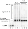
Figure 1
Figure 1
Product distributions from the histone transfer reaction onto the S1 duplex. Lanes 1,2: free DNA; lanes 3–5: after completion of transfer reactions at 4 °C′ lanes 6–8: after heat equilibration at 45°C/2h. The average length of donor chromatin in lanes 3, 6: 750 bp; in lanes 4, 7: 450 bp; in lanes 4,8: 250 bp.
Figure 2
Figure 2
Autoradiographs of footprinting experiments. (a) The template strands of the duplexes were 5′-labeled with 32P. The positions of the platinum adduct are highlighted by red arrows. Lane 1: S1, lane 2: S1-Pt, lane 3: S2, lane 4: S2-Pt, lane 5: nS1, lane 6: nS1-Pt, lane 7: nS2, lane 8: nS2-Pt, lane M1: Maxam-Gilbert A/G reaction of S1, lane M2: Maxam-Gilbert A/G reaction of S1-Pt, lane M3: Maxam-Gilbert A/G reaction of S2, lane M4: Maxam-Gilbert A/G reaction of S2- Pt; (b) the coding strands of the duplexes were 5′-labeled. The positions of the d(CpApC) trinucleotide opposite the platinum adduct are highlighted by red arrows. Lane 1: S1, lane 2: S1-Pt, lane 3: S2, lane 4: S2-Pt, lane 5: nS1, lane 6: nS1-Pt, lane 7: nS2, lane 8: nS2-Pt, lane M1: Maxam-Gilbert A/G reaction of S1, lane M2: Maxam-Gilbert A/G reaction of S1-Pt, lane M3: Maxam-Gilbert A/G reaction of S2, lane M4: Maxam-Gilbert A/G reaction of S2-Pt.
Figure 3
Figure 3
Superimposition of the cleavage curves of (a) unplatinated and (b) platinated nucleosomes. (a) The DNA sequences of the unplatinated nucleosomes nS1 (blue) and nS2 (green) are identical except for the fact that a d(GpTpG) trinucleotide in the bottom strand was shifted by 5 bp. The corresponding cleavage curves are out of phase by 1–2 bp. (b) The platinated nuclesosomes nS1-Pt (red) and nS2-Pt (orange) have identical sequences except for the fact that the 1,3-d(GpTpG) cisplatin adduct, symbolized by a black rectangle, was shifted by 5 bp. The corresponding cleavage curves are out of phase by 3–5 bp. The phasing difference is almost 180° in the vicinity of the cisplatin adduct.
Figure 4
Figure 4
The cleavage curves reveal the rotational setting of the nucleosomal duplexes nS1, nS1-Pt, nS2, and nS2-Pt in the nucleosome. Gray bars indicate areas in which the minor groove is exposed to the solvent and the major groove faces inward to the nucleosome. (a) The major groove of the unplatinated d(GpTpG) trinucleotide of S1 faces inward, (b) the major groove of the unplatinated d(GpTpG) site of nS2 faces sideways, (c) and (d), the major groove of the platinated d(GpTpG) sites faces in both cases inward. Cisplatin, which binds to the purine bases in the major groove of DNA, is in both cases buried inside the nucleosome, inaccessible to the solvent.
Figure 5
Figure 5
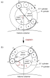
Schematic projection of the d(GpTpG) trinucleotide (position 91.93) of the nucleosomes (a) nS2 and (b) nS2-Pt along the central axis of the nucleosomal DNA. The two concentric circles indicate expected locations of the P- and C1′-atoms in the DNA duplex. The G:C base pairs are symbolized by thick lines. (a) The major groove of the unplatinated d(GpTpG) trinucleotide points sideways (ξ ≈ 90°) and its backbone faces directly to the solvent. (b) The major groove of the d(GpTpG) trinucleotide and the platinum adduct point toward the histone octamer (ξ ≈ 0°) and its minor grove faces directly toward the solvent.
Figure 6
Figure 6
Superimposition of the cleavage curves of the unplatinated nucleosome nS1 and the corresponding platinated nucleosome nS1-Pt shows that the cisplatin adduct unwinds the DNA strand on the nucleosome by approximately 1 bp.
Figure 7
Figure 7
Helical periodicities of nucleosomal DNA strands. (a) n1S (blue) and n1S-Pt (red), (b), n2S (green) and n2S-Pt (orange) superimposed. The positions of the platinum cross-links are indicated by black rectangles and the pseudodyad axes by arrows. Comparison of platinated and unplatinated nucleosomes reveals a local underwinding and overwinding in vicinity of the cisplatin adduct.
Scheme 1
Scheme 1
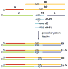
Construction of site-specifically platinated nucleosome positioning sequences.

Similar articles

Cited by

References

    1. Wong E, Giandomenico CM. Chem Rev. 1999;99:2451–2466. - PubMed
    1. Jamieson ER, Lippard SJ. Chem Rev. 1999;99:2467–2498. - PubMed
    1. Wang D, Lippard SJ. Nat Rev Drug Discovery. 2005;4:307–319. - PubMed
    1. Blommaert FA, van Dijk-Knijnenburg HCM, Dijt FJ, den Engelse L, Baan RA, Berends F, Fichtinger-Schepman AMJ. Biochemistry. 1995;34:8474–8480. - PubMed
    1. Lippard SJ, Hoeschele JD. Proc Natl Acad Sci U S A. 1979;76:6091–6095. - PMC - PubMed

Publication types