Review
. 2007 Apr 14:4:27.
doi: 10.1186/1742-4690-4-27.

Centrosome and retroviruses: the dangerous liaisons

Affiliations
Review

Centrosome and retroviruses: the dangerous liaisons

Philippe V Afonso et al. Retrovirology. .

Abstract

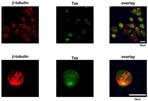
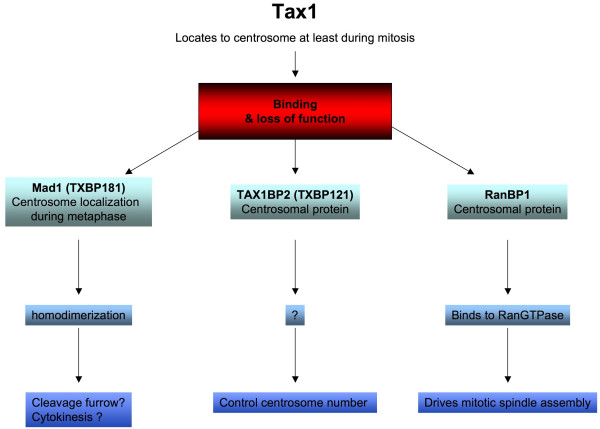
Centrosomes are the major microtubule organizing structures in vertebrate cells. They localize in close proximity to the nucleus for the duration of interphase and play major roles in numerous cell functions. Consequently, any deficiency in centrosome function or number may lead to genetic instability. Several viruses including retroviruses such as, Foamy Virus, HIV-1, JSRV, M-PMV and HTLV-1 have been shown to hamper centrosome functions for their own profit, but the outcomes are very different. Foamy viruses, HIV-1, JSRV, M-PMV and HTLV-1 use the cellular machinery to traffic towards the centrosome during early and/or late stages of the infection. In addition HIV-1 Vpr protein alters the cell-cycle regulation by hijacking centrosome functions. Enthrallingly, HTLV-1 Tax expression also targets the functions of the centrosome, and this event is correlated with centrosome amplification, aneuploidy and transformation.


Figures

Figure 1
Figure 1
The centrosome duplication cycle. Adapted from [3, 14, 107]. Centrosome duplication starts at the G1/S transition with the separation of the paired centrioles. Procentrioles form near the proximal ends of each pre-existing centriole. Procentriole formation is completed during S phase. During mitosis the two centrosomes are present and form the mitotic spindle poles. Each cell inherits one centrosome after the completion of cytokinesis.
Figure 2
Figure 2
Retroviruses target the MTOC during the early and/or the late phase of the viral replication cycle. Retroviruses enter into the host cell mainly by receptor-mediated fusion of the viral envelope with the plasma membrane (1). After crossing the actin cortex, the viral core is released into the cytoplasm where it undergoes a process of uncoating during which the viral genomic RNA (black) is reverse transcribed into a double-strand linear DNA copy (red) (2). Incoming viral cores en route to the nucleus reach the MTOC by using the molecular motor complexes to traffic along the MTs (3). In the nucleus the viral DNA genome is stably integrated into the host cell chromosome (4). The integrated viral DNA or provirus is the template for the synthesis of the viral mRNAs (5) which are transported in the cytoplasm and translated to produce the viral Gag polyproteins and the viral envelope glycoproteins(6). Newly synthesized Gag proteins and the viral genomic RNA converge to the MTOC where encapsidation and assembly initiate (7a). At this late stage, FVs are characterized by a second reverse transcription event (7b). By trafficking along the MT network assembling viral particles reach the plasma or endosomal membranes where budding occurs (8a and b). Finally, for most retroviruses, a process known as maturation is necessary for the generation of infectious viruses able to begin a new round of infection (9).
Figure 3
Figure 3
Vpr induces a G2 cell cycle arrest and eventually apoptosis. (A) In uninfected cells Polo like kinase-1 (Plk1) activates the Mitosis Promoting Factor (MPF) both directly and indirectly by inhibiting the Wee1 and the Mut1 kinases. Cdc25 proteins are also involved in MPF activation via a positive retroactive loop. In normal cells, MPF activation leads to mitosis; in non-viable cells, mitosis acts as a checkpoint and cells die by apoptosis. (B) In HIV-1 infected cells, Vpr expression induces the relocalization of Plk1 and form a ternary complex with 14-3-3 and Cdc25. MPF cannot be activated, and consequently cells arrest in G2 phase. Non-viable (polyploid) cells transiently accumulate. If the cell-cycle arrest is prolonged, cells die by necrosis.
Figure 4
Figure 4
In naturally infected T cells a substantial fraction of Tax co-localizes with the MTOC. Images of CD4+ T cells naturally infected with HTLV-1 obtained from a TSP/HAM patient. Cells were stained with anti-Tax mAb Lt-4 (green) and monoclonal anti-β Tubulin-Cy3 antibody (red). The view represents a projection (XY axis) of adjacent confocal sections. Scale bar = 10 μM.
Figure 5
Figure 5
Pleiotropic actions of Tax-1. Tax-1 interacts and impairs the function of at least with 3 different centrosomal proteins (Mad1, TAX1BP2 and RanBP1) which participate in the control of mitosis.

Similar articles

Cited by

References

    1. Boveri T. Über merhrpolige Mitosen als Mittel zur analyse des zellkerns. Verh D Phys Med Ges Würzburg NF. 1902;35:67–90.
    1. Andersen JS, Wilkinson CJ, Mayor T, Mortensen P, Nigg EA, Mann M. Proteomic characterization of the human centrosome by protein correlation profiling. Nature. 2003;426:570–574. doi: 10.1038/nature02166. - DOI - PubMed
    1. Fukasawa K. Centrosome amplification, chromosome instability and cancer development. Cancer Lett. 2005;230:6–19. doi: 10.1016/j.canlet.2004.12.028. - DOI - PubMed
    1. Burkhard P, Stetefeld J, Strelkov SV. Coiled coils: a highly versatile protein folding motif. Trends Cell Biol. 2001;11:82–88. doi: 10.1016/S0962-8924(00)01898-5. - DOI - PubMed
    1. Tsou MF, Stearns T. Controlling centrosome number: licenses and blocks. Curr Opin Cell Biol. 2006;18:74–78. doi: 10.1016/j.ceb.2005.12.008. - DOI - PubMed

Publication types

LinkOut - more resources