. 2007 Apr 16;8(1):31.
doi: 10.1186/1465-9921-8-31.

Proteinase-activated receptor 4 stimulation-induced epithelial-mesenchymal transition in alveolar epithelial cells

Affiliations

Proteinase-activated receptor 4 stimulation-induced epithelial-mesenchymal transition in alveolar epithelial cells

Seijitsu Ando et al. Respir Res. .

Abstract

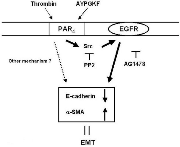
Background: Proteinase-activated receptors (PARs; PAR1-4) that can be activated by serine proteinases such as thrombin and neutrophil catepsin G are known to contribute to the pathogenesis of various pulmonary diseases including fibrosis. Among these PARs, especially PAR4, a newly identified subtype, is highly expressed in the lung. Here, we examined whether PAR4 stimulation plays a role in the formation of fibrotic response in the lung, through alveolar epithelial-mesenchymal transition (EMT) which contributes to the increase in myofibroblast population.

Methods: EMT was assessed by measuring the changes in each specific cell markers, E-cadherin for epithelial cell, alpha-smooth muscle actin (alpha-SMA) for myofibroblast, using primary cultured mouse alveolar epithelial cells and human lung carcinoma-derived alveolar epithelial cell line (A549 cells).

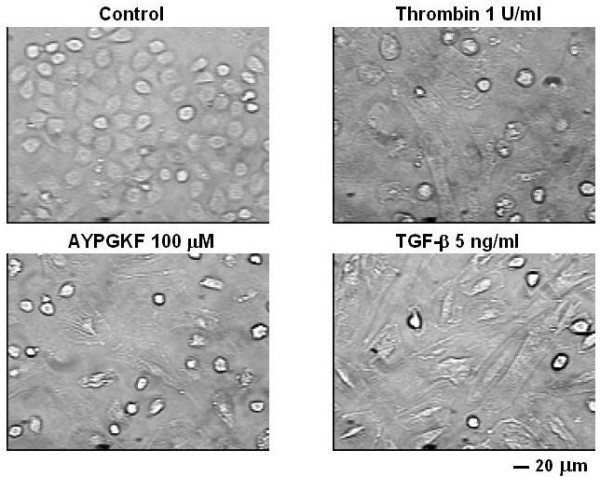
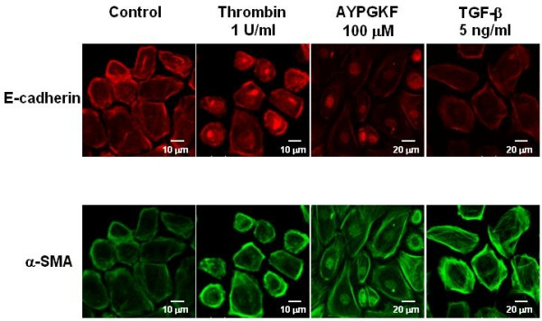
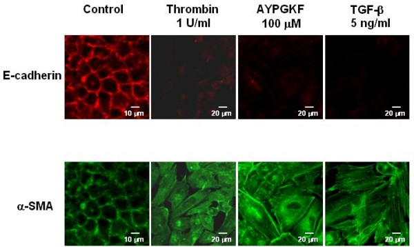
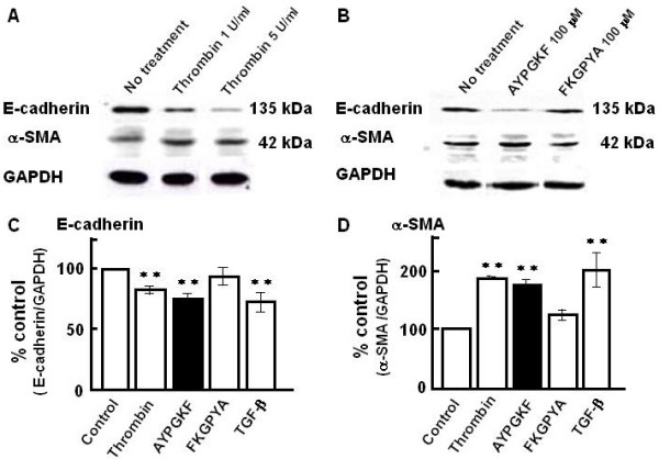
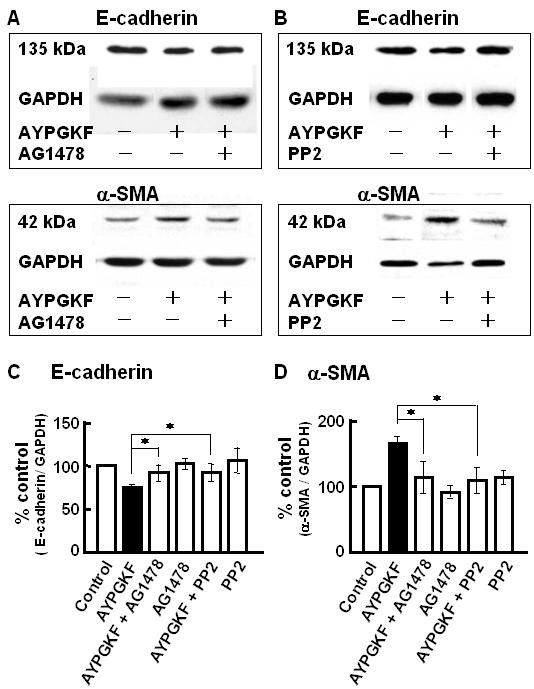
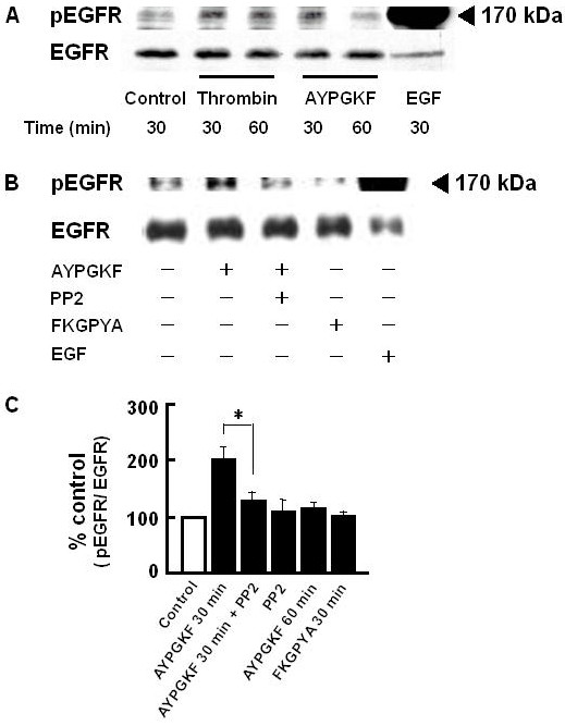
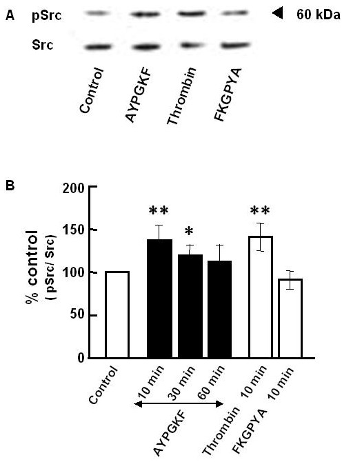
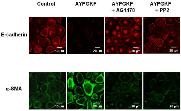
Results: Stimulation of PAR with thrombin (1 U/ml) or a synthetic PAR4 agonist peptide (AYPGKF-NH2, 100 muM) for 72 h induced morphological changes from cobblestone-like structure to elongated shape in primary cultured alveolar epithelial cells and A549 cells. In immunocytochemical analyses of these cells, such PAR4 stimulation decreased E-cadherin-like immunoreactivity and increased alpha-SMA-like immunoreactivity, as observed with a typical EMT-inducer, tumor growth factor-beta (TGF-beta). Western blot analyses of PAR4-stimulated A549 cells also showed similar changes in expression of these EMT-related marker proteins. Such PAR4-mediated changes were attenuated by inhibitors of epidermal growth factor receptor (EGFR) kinase and Src. PAR4-mediated morphological changes in primary cultured alveolar epithelial cells were reduced in the presence of these inhibitors. PAR4 stimulation increased tyrosine phosphorylated EGFR or tyrosine phosphorylated Src level in A549 cells, and the former response being inhibited by Src inhibitor.

Conclusion: PAR4 stimulation of alveolar epithelial cells induced epithelial-mesenchymal transition (EMT) as monitored by cell shapes, and epithelial or myofibroblast marker at least partly through EGFR transactivation via receptor-linked Src activation.


Figures

Figure 1
Figure 1
Typical phase contrast images of primary cultured mouse alveolar epithelial cells. Epithelial cells were treated for 72 h without agonist (control) or with thrombin (1 U/ml), PAR4 agonist peptide (AYPGKF-NH2, 100 μM) or TGF-β (5 ng/ml).
Figure 2
Figure 2
Phenotypic changes in primary cultured alveolar epithelial cells stimulated with PAR4 agonists or TGF-β. Immunofluorescence images for a specific marker for epithelial cell (E-cadherin; rhodamine red, upper panel) or myofibroblast (α-SMA; FITC green, lower panel) captured with confocal lasar microscopy. Cells were treated with or without (control) various agonists (thrombin, AYPGKF-NH2 or TGF-β) for 72 h, and stained for E-cadherin or α-SMA using specific antibodies as described in Method section.
Figure 3
Figure 3
Phenotypic changes in A549 cells stimulated with PAR4 agonists or TGF-β. Immunofluorescence images for a specific marker for epithelial cell (E-cadherin; rhodamine red, upper panel) or myofibroblast (α-SMA; FITC green, lower panel) captured with confocal lasar microscopy. Cells were treated with or without (control) various agonists (thrombin, AYPGKF-NH2 or TGF-β) for 72 h, and stained for E-cadherin or α-SMA using specific antibodies as described in Method section.
Figure 4
Figure 4
Western blot analyses of E-cadherin and α-SMA in A549 epithelial cells stimulated with PAR4 agonists and TGF-β. (A, B) Representative Western blots of E-cadherin and α-SMA in A549 cells treated for 96 h with thrombin (A; 1 or 5 U/ml) or synthetic peptides (B; AYPGKF-NH2 or FKGPYA-NH2; 100 μM). (C, D) Summarized densitometric data. Effects of PAR4 agonists (thrombin; 1 U/ml and AYPGKF-NH2; 100 μM), FKGPYA-NH2 (100 μM) or TGF-β (5 ng/ml) on the expressions of E-cadherin (C) and α-SMA (D). Results are based on densitometric analyses of the ratio of each marker protein to GAPDH (as internal standard). Each bar represents the mean ± S.E.M. from 4–12 preparations. **P < 0.01 compared with the control.
Figure 5
Figure 5
Effects of inhibitors of EGFR tyrosine kinase and Src tyrosine kinase on PAR4 agonist-induced changes in EMT-related marker protein expression in A549 cells. A549 cells were pretreatd with or without 30 nM AG1478 or 300 nM PP2 for 30 min, and then stimulated with AYPGKF-NH2 (100 μM) for 96 h followed by immunoblotting with specific antibodies for E-cadherin, α-SMA. (A, B) Representative Western blots for the effect of AG1478 (A) or PP2 (B) on the expression of E-cadherin and α-SMA. (C, D) Summarized densitometric data. Each bar represents the mean ± S.E.M. for 4–12 preparations. *P < 0.05 compared with AYPGKF-NH2 alone.
Figure 6
Figure 6
PAR4 agonist-induced EGFR phosphorylation in A549 cells. A549 cells were pretreated with or without 300 nM PP2 for 30 min, and then stimulated with AYPGKF-NH2 (100 μM) for 30–60 min followed by immunoblotting with specific antibodies for the Y1173 phosphorylated EGFR and total EGFR protein. Control; without drug treatment. (A) Representative time course of EGFR phosphorylation induced by thrombin (1 U/ml) or AYPGKF-NH2 (100 μM). As a positive control, cells were treated with EGF (10 ng/ml) for 30 min. (B) Representative Western blot after treatment with or without (control) various agents (AYPGKF-NH2, AYPGKF-NH2+PP2, 100 μM FKGPYA-NH2 or EGF) for 30 min. (C) Summarized densitometric data. Protein concentrations used for separation was 20 μg/lane (thrombin or AYPGKF-NH2 or FKGPYA-NH2 treated sample) or 10 μg/lane (EGF treated sample). Each bar represents the mean ± S.E.M. for 4–6 preparations. *P < 0.05 compared with AYPGKF-NH2 alone.
Figure 7
Figure 7
Western blot analyses of Src phosphorylation in A549 cells. A549 cells were treated without (control) or with AYPGKF-NH2 (100 μM) for 10, 30 or 60 min or with thrombin (1 U/ml, 10 min) or with FKGPYA-NH2 (100 μM, 10 min), and then subjected to immunoblotting with specific antibodies for the Y418 phosphorylated Src and total Src protein. (A) Representative Western blot. Stimulation time was 10 min. (B) Summarized densitometric data. Each bar represents the mean ± S.E.M. for 4–6 preparations. **P < 0.01, *P < 0.05 compared with the control.
Figure 8
Figure 8
Effects of AG1478 and PP2 on PAR4 agonist induced phenotypic changes in primary cultured alveolar epithelial cells. Immunofluorescence images for a specific marker for epithelial cell (E-cadherin; rhodamine red, upper panel) or myofibroblast (α-SMA; FITC green, lower panel) captured with confocal lasar microscopy. Cells were treated with or without (control) AYPGKF-NH2 (100 μM) for 72 h in the presence or absence of each inhibitor (30 nM AG1478 or 300 nM PP2), and stained using each specific antibody as described in Method section.
Figure 9
Figure 9
A schematic illustration summarizing the mechanisms for PAR4-mediated EMT in alveolar epithelial cell. Bold line: a new pathway proposed in this study. Dotted line: other mechanisms.

References

    1. Lan RS, Stewart GA, Henry PJ. Role of protease-activated receptors in airway function: a target for therapeutic intervention? Pharmacol Ther. 2002;95:239–257. doi: 10.1016/S0163-7258(02)00237-1. - DOI - PubMed
    1. Macfarlane SR, Seatter MJ, Kanke T, Hunter GD, Plevin R. Protease-activated receptors. Phamacol Rev. 2001;53:245–282. - PubMed
    1. Howell DC, Johns RH, Lasky JA, Shan B, Scotton CJ, Laurent GJ, Chambers RC. Absence of protease-activated receptor-1 signaling affords protection from bleomycin-induced lung inflammation and fibrosis. Am J Pathol. 2005;166:1353–1365. - PMC - PubMed
    1. Yagi Y, Otani H, Ando S, Oshiro A, Kawai K, Nishikawa H, Araki H, Fukuhara S, Inagaki C. Involvement of Rho signaling in PAR2-mediated regulation of neutrophil adhesion to lung epithelial cells. Eur J Pharmacol. 2006;536:19–27. doi: 10.1016/j.ejphar.2006.02.024. - DOI - PubMed
    1. Hernández-Rodriguez NA, Cambrey AD, Harrison NK, Chambers RC, Gray AJ, Southcott AM, duBois RM, Black CM, Scully MF, McAnulty RJ, Laurent GJ. Role of thrombin in pulmonary fibrosis. Lancet. 1995;346:1071–1073. doi: 10.1016/S0140-6736(95)91744-6. - DOI - PubMed

Publication types

MeSH terms