. 2007 Jun 1;306(1):121-33.
doi: 10.1016/j.ydbio.2007.03.011. Epub 2007 Mar 13.

Wnt5a functions in planar cell polarity regulation in mice

Affiliations

Wnt5a functions in planar cell polarity regulation in mice

Dong Qian et al. Dev Biol. .

Abstract

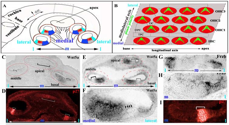
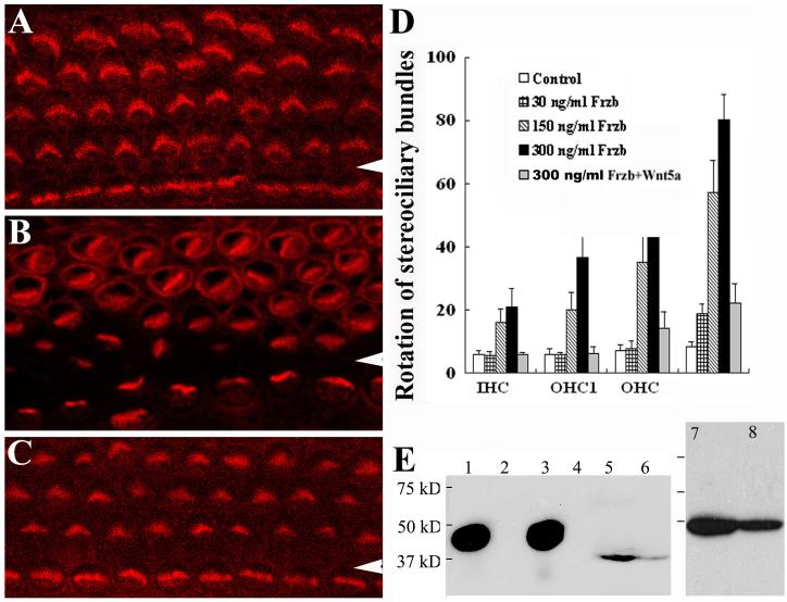
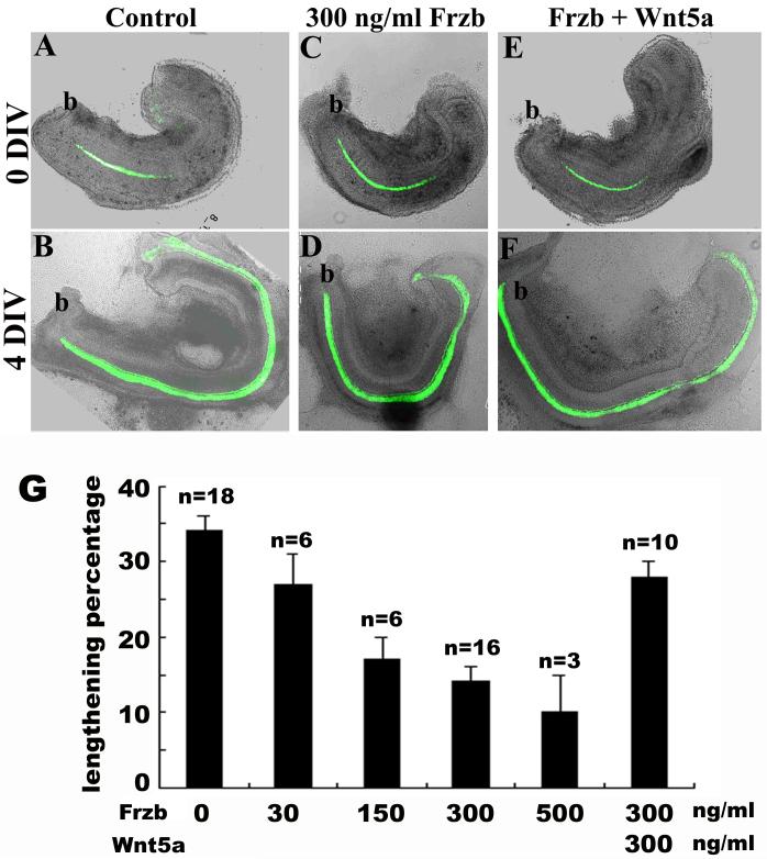
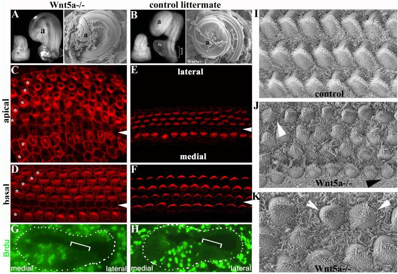
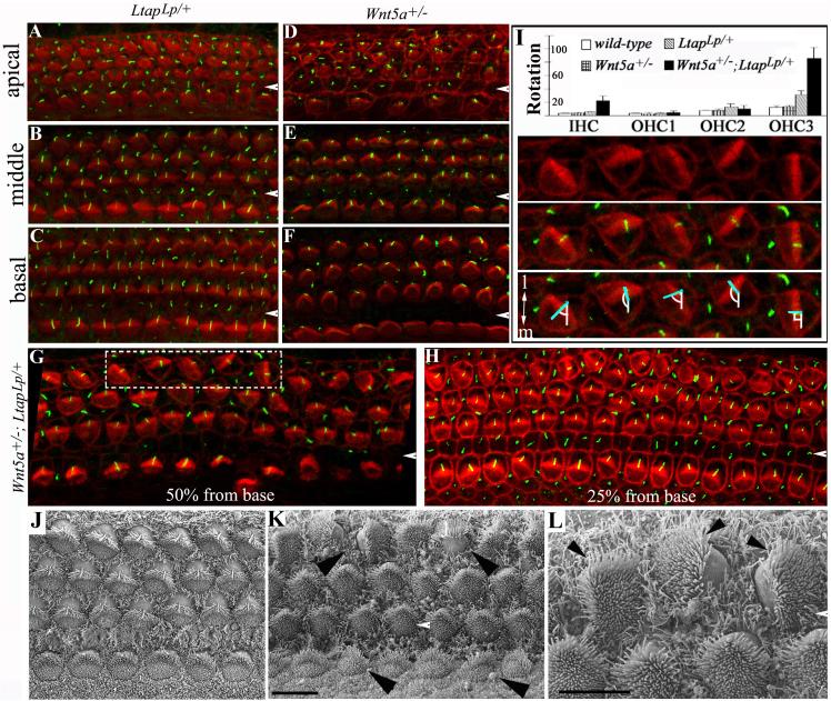
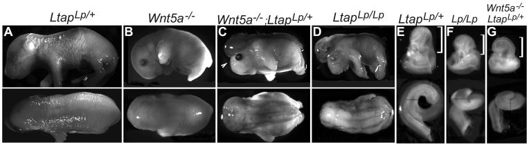
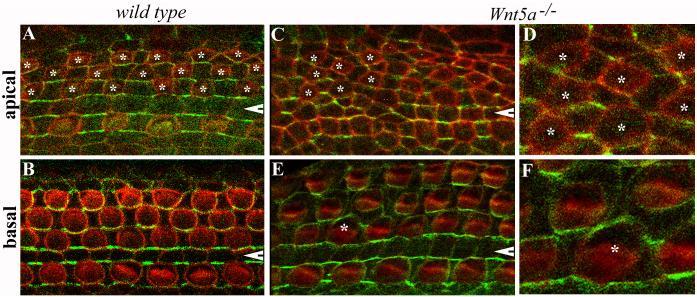
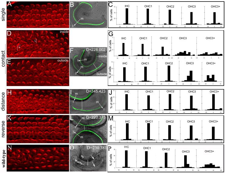
Planar cell polarity (PCP) refers to the polarization of cells within the plane of a cell sheet. A distinctive epithelial PCP in vertebrates is the uniform orientation of stereociliary bundles of the sensory hair cells in the mammalian cochlea. In addition to establishing epithelial PCP, planar polarization is also required for convergent extension (CE); a polarized cellular movement that occurs during neural tube closure and cochlear extension. Studies in Drosophila and vertebrates have revealed a conserved PCP pathway, including Frizzled (Fz) receptors. Here we use the cochlea as a model system to explore the involvement of known ligands of Fz, Wnt morphogens, in PCP regulation. We show that Wnt5a forms a reciprocal expression pattern with a Wnt antagonist, the secreted frizzled-related protein 3 (Sfrp3 or Frzb), along the axis of planar polarization in the cochlear epithelium. We further demonstrate that Wnt5a antagonizes Frzb in regulating cochlear extension and stereociliary bundle orientation in vitro, and that Wnt5a(-/-) animals have a shortened and widened cochlea. Finally, we show that Wnt5a is required for proper subcellular distribution of a PCP protein, Ltap/Vangl2, and that Wnt5a interacts genetically with Ltap/Vangl2 for uniform orientation of stereocilia, cochlear extension, and neural tube closure. Together, these findings demonstrate that Wnt5a functions in PCP regulation in mice.


Figures

Fig. 1
Fig. 1. Reciprocal expression of Wnt5a and Frzb along the mediolateral axis of the cochlea
(A): Diagram of a cross section of the cochlear duct at different regions along the longitudinal axis (outlined by red circles). The IHCs are localized toward the center, or medial; the three rows of OHCs are at the periphery, or lateral region. The differentiation gradient of the organ of Corti is designated by arrows near base of the cochlea (A). The dark and light blue regions correspond, respectively, to the Wnt5a- and Frzb-expressing domains. (B): Stereocilia are uniformly oriented along the mediolateral axis illustrated in this diagram of a whole mount of the organ of Corti. (C,D): At E14.5, Wnt5a mRNA was detected in the region medial (C) to the developing organ of Corti (indicated by brackets) at the basal region of the cochlea. The expression of p27/Kip1 (D, red) marked distinctively the developing organ of Corti at this stage. In the middle and apical regions of the cochlea, Wnt5a expression was not detected. (E,F): At E16.5, Wnt5a is expressed in the region medial to the organ of Corti (indicated with brackets) along the length of the cochlear duct. (F) is a higher magnification view of the middle cochlear cross section of (E). The IHC and OHC are indicated by an arrowhead and arrows, respectively. (G-I): At E14.5, Frzb is expressed in the regions lateral (H, I) to the developing organ of Corti (indicated by brackets) marked by p27/Kip1 expression (J). (I): a higher magnification view of (H). m: medial. l: lateral.
Fig. 2
Fig. 2. Wnt5a and Frzb interact in vitro for stereociliary bundle orientation
(A-D): The cochleae from wild-type E14.5 embryos undergo differentiation in culture. Stereocilia were normally oriented in the control medium as shown by phalloidin staining (A). The addition of Frzb to the medium affected stereociliary bundle orientation (B), but this effect was suppressed by pre-incubation with Wnt5a (C). Arrowheads indicate the pillar cell region that separates the IHCs from OHCs. Stereociliary bundle misorientation in the culture was quantified (D). The OHC3 and additional rows of OHCs in culture are quantified as a single group, OHC3+. The effect of Frzb on stereociliary bundle orientation ([Frzb]> 150 ng/ml, p<0.001) and the rescue by Wnt5a pre-incubation (p<0.000) were statistically significant. (E): Western blots with an antibody against Wnt5a. Lane 1: purified recombinant Wnt5a (R&D); lanes 2 and 4: media from the control cell line; lane 3: media from Wnt5a-expressing cells (Wnt5a-CM); lanes 5, 6, 8: cochlear extracts; lane 7: brain extracts.
Fig. 3
Fig. 3. Wnt5a suppressed the effect of Frzb on cochlear extension in vitro
(A-F): The extension of the wild-type cochlea in culture was reduced in the presence of Frzb (A-D). This reduced extension was partially suppressed by pre-incubation with Wnt5a (E,F). Green signals are GFP expressed under the control of Math1 enhancers marking the hair cells. At the beginning of culture, only a single row of IHCs were differentiated near the base of the cochlea (A, C, E). b: base of the cochlea. (G): Quantification of cochlear extension in vitro. The effect of Frzb on cochlear extension ([Frzb]>150 ng/ml, p<0.001) and the rescue by Wnt5a pre-incubation (p=0.001) were statistically significant.
Fig. 4
Fig. 4. Wnt5a null animals show characteristic CE defects in the cochlea
(A-B): Cochleae from E18.5 Wnt5a−/− embryos (A) were shorter than those from control littermates (B) as shown in both dissection (left) and SEM (right) images. Dashes outline the curvatures of the cochlear ducts. Sa: vestibular saccule attached to the base of the cochlea. (C-F) The organ of Corti from an E18.5 Wnt5a−/− embryo (C-D) stained with phalloidin showed additional rows of OHCs in both basal and apical regions compared with a control littermate (E,F). Asterisks mark each row of OHCs in the Wnt5a−/− sample. Arrowheads indicate the pillar cell region. The confocal plane for the Wnt5a−/− sample was close to the apical cortex of the cells in the organ of Corti, and therefore the phalloidin staining for cell-cell junctions in the Wnt5a−/− sample (C,D) appeared to be more intense than the staining in the control sample (E,F). (G-H): BrdU-injected E18.5 cochlear sections from Wnt5a−/− (G) and control (H, Wnt5a+/−) littermates stained with BrdU antibody (green) to reveal dividing cells. Brackets indicate the organ of Corti. Dashes dots outline the cochlear ducts. No statistic difference in the number of BrdU+ cells was detected in comparable cochlear sections from Wnt5a−/− and control embryos. (I-K): In contrast to normal polarity of the stereocilia across the organ of Corti in the control littermate (I), the organ of Corti from Wnt5a−/− animals showed limited misorientation of stereocilia with varying degrees (J, K). The white arrowheads indicate a rotated outer hair cell (J) and a pair of outer hair cells facing each other (K). The black arrowhead indicates a rotated inner hair cell (J).
Fig. 5
Fig. 5. Genetic interaction of Wnt5a and Ltap/Vangl2 in establishing uniform orientation of stereocilia
(A-H): The cochlear ducts from E18.5 LtapLp/+ (A-C), Wnt5a+/− (D-F), and Wnt5a+/−; LtapLp/+ (G-H) littermates stained with phalloidin (red) and acetylated α-tubulin (green). The acetylated α-tubulin antibody labels the kinocilia located at the vertices of the stereocilia and phalloidin labels actin in stereocilia. In contrast to the heterozygous LtapLp/+ (A-C) and Wnt5a+/− (D-F) embryos, the Wnt5a+/−; LtapLp/+ double-heterozygous littermates (G-H) had misoriented stereocilia in the third row of OHCs. The white arrowheads indicate the pillar cell region separating the IHCs from OHCs. (I): The orientation of stereocilia in Wnt5a+/−; LtapLp/+ (n= 16) mice was quantified and compared to that of LtapLp/+ (n = 4), Wnt5a+/− (n = 4) and wild-type (n = 4) animals. The quantification of stereociliary orientation (I) is illustrated by hair cells from the dashed box in (G), which is shown in the panels below the histogram in (I) with the top panel showing phalloidin labeling alone, the middle panel showing phalloidin plus acetylated atubulin, and bottom panel showing the measurement of the rotation. The angle formed between the mediolateral axis (I, bottom panel, white line) and the line bisecting the “V”-shaped stereocilia (I, bottom panel, blue line) was measured and plotted in the histogram. (J-L): SEM images of the organ of Corti from E17.5 LtapLp/+ (J) and Wnt5a+/−; LtapLp/+ (K-L) embryos. Arrowheads in (K) mark two OHCs with an abnormal 90° or 180° rotation and two IHCs with minor degrees of rotation. The misalignment of stereocilia (L, arrowheads) is more clearly seen at high magnification (L, higher magnification of the top left corner of K). Scale bars: 10 μm (J-K) and 6 μm (L).
Fig. 6
Fig. 6. Genetic interaction of Wnt5a and Ltap for neurulation and cochlear lengthening
(A-D): LtapLp/+ (n>100) (A) and Wnt5a−/− (B, 95%, n = 26) mice had normal neural tube closure. Wnt5a−/−;LtapLp/+ (C, 100%, n = 4) and LtapLp/Lp embryos (D, 100%) show craniorachischisis observed in all the known PCP mutants. The arrowhead in (C) indicates the open- eyelid phenotype observed in several PCP mutants. (E-G): Inner ears and dissected cochlear ducts (below) from LtapLp/+ (E), LtapLp/Lp (F) and Wnt5a−/−;LtapLp/+ embryos (G). The brackets indicate the cochlear portion. The lines were drawn from the medial to the lateral sides of the cochlear ducts in the apical region to mark the width of the cochlear ducts.
Fig. 7
Fig. 7. Aberrant cellular geometries and distribution of Ltap in Wnt5a−/− mice
(A-F): Whole mount of the organ of Corti from E18.5 wild-type (A-B) and Wnt5a−/− (C-F) mice carrying BACLtap-GFP. Hair cells were visualized by phalloidin staining (red). Ltap-GFP (green) was seen at the medial boundaries between hair cells and supporting cells in wild-type samples (A, B). In Wnt5a−/− mice, the localization of Ltap-GFP in the base region appeared normal on the medial side of hair cells (E), except around one hair cell (E, marked by an asterisk). In the apical region, cellular geometry was altered in Wnt5a−/− mice, and so was the localization of Ltap-GFP (C). OHCs in the apical region are marked with asterisks (A, C). White arrowheads indicate the pillar cell region. (D) and (F) are higher magnification images for the regions where hair cells are marked by asterisks in (C) and (E), respectively.
Fig. 8
Fig. 8. Potential instructive cues along the mediolateral axis of the cochlea
(A-P): The orientation of stereociliary bundles was visualized with phalloidin staining (A,D,E,H,K,N) in wild-type cochleae cultured singly (A,B), in parallel closely (D-F), in parallel distantly (H,I), in anti-parallel (K,L), and between closely parallel Wnt5a−/− and wild-type cochleae (N,P) The distribution of stereociliary bundle orientation in each row of hair cells cochleae cultured singly (C, n=11), in closely parallel (G, n=4), in distantly parallel (J, n=4), in anti-parallel (M, n=4), and between Wnt5a−/− and wild-type cochleae (P, N=3) was quantified and plotted on the histograms. The angles formed between the mediolateral axis (D, white lines) and the lines bisecting the “V”-shaped stereocilia (D, blue lines) were used for quantifications as described in Methods. This angle is close to 0 in normally oriented stereocilia. As diagramed in (D), the angles formed at the right or the left sides of the mediolateral axis were designated to be positive or negative, respectively. The lines (F,I,L,O) and arrows (B,F,I,L,O) indicate the distance between the two cochleae and the orientation of the mediolateral axes of the cochleae, respectively. The green signal (B, F, I, L) is from GFP expressed under Math1 enhancers and marks the hair cells. In the grafts between Wnt5a−/− and wild-type cochleae (O), GFP was not present and the cochleae were outlined by dashed lines. D: units of the distance between the two cochleae cultured together using NIH ImageJ (F, I, L,O); m: the medial side of the cochlea; l: the lateral side of the cochlea.

References

    1. Amonlirdviman K, Khare NA, Tree DR, Chen WS, Axelrod JD, Tomlin CJ. Mathematical modeling of planar cell polarity to understand domineering nonautonomy. Science. 2005;307:423–6. - PubMed
    1. Badea TC, Wang Y, Nathans J. A noninvasive genetic/pharmacologic strategy for visualizing cell morphology and clonal relationships in the mouse. J Neurosci. 2003;23:2314–22. - PMC - PubMed
    1. Bhanot P, Brink M, Samos CH, Hsieh JC, Wang Y, Macke JP, Andrew D, Nathans J, Nusse R. A new member of the frizzled family from Drosophila functions as a Wingless receptor. Nature. 1996;382:225–30. - PubMed
    1. Blankenship JT, Backovic ST, Sanny JS, Weitz O, Zallen JA. Multicellular rosette formation links planar cell polarity to tissue morphogenesis. Dev Cell. 2006;11:459–70. - PubMed
    1. Chen P, Johnson JE, Zoghbi HY, Segil N. The role of Math1 in inner ear development: Uncoupling the establishment of the sensory primordium from hair cell fate determination. Development. 2002;129:2495–505. - PubMed

Publication types