Review
. 2007 May;32(5):225-34.
doi: 10.1016/j.tibs.2007.03.002. Epub 2007 Apr 12.

Bacterial response regulators: versatile regulatory strategies from common domains

Affiliations
Review

Bacterial response regulators: versatile regulatory strategies from common domains

Rong Gao et al. Trends Biochem Sci. 2007 May.

Abstract

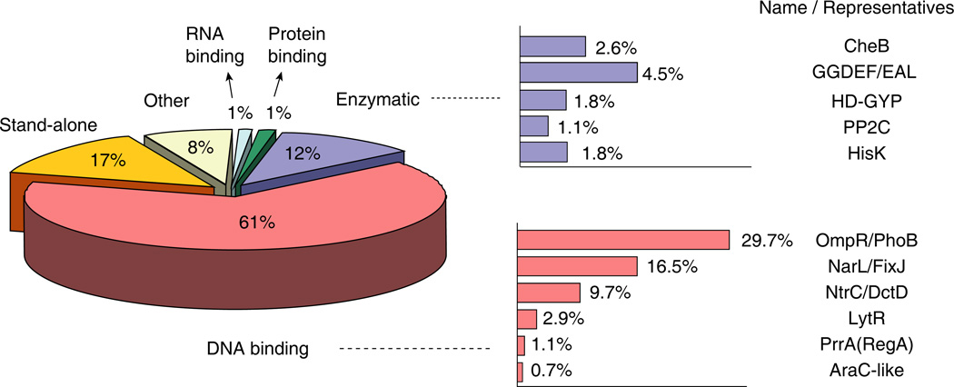
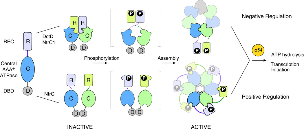
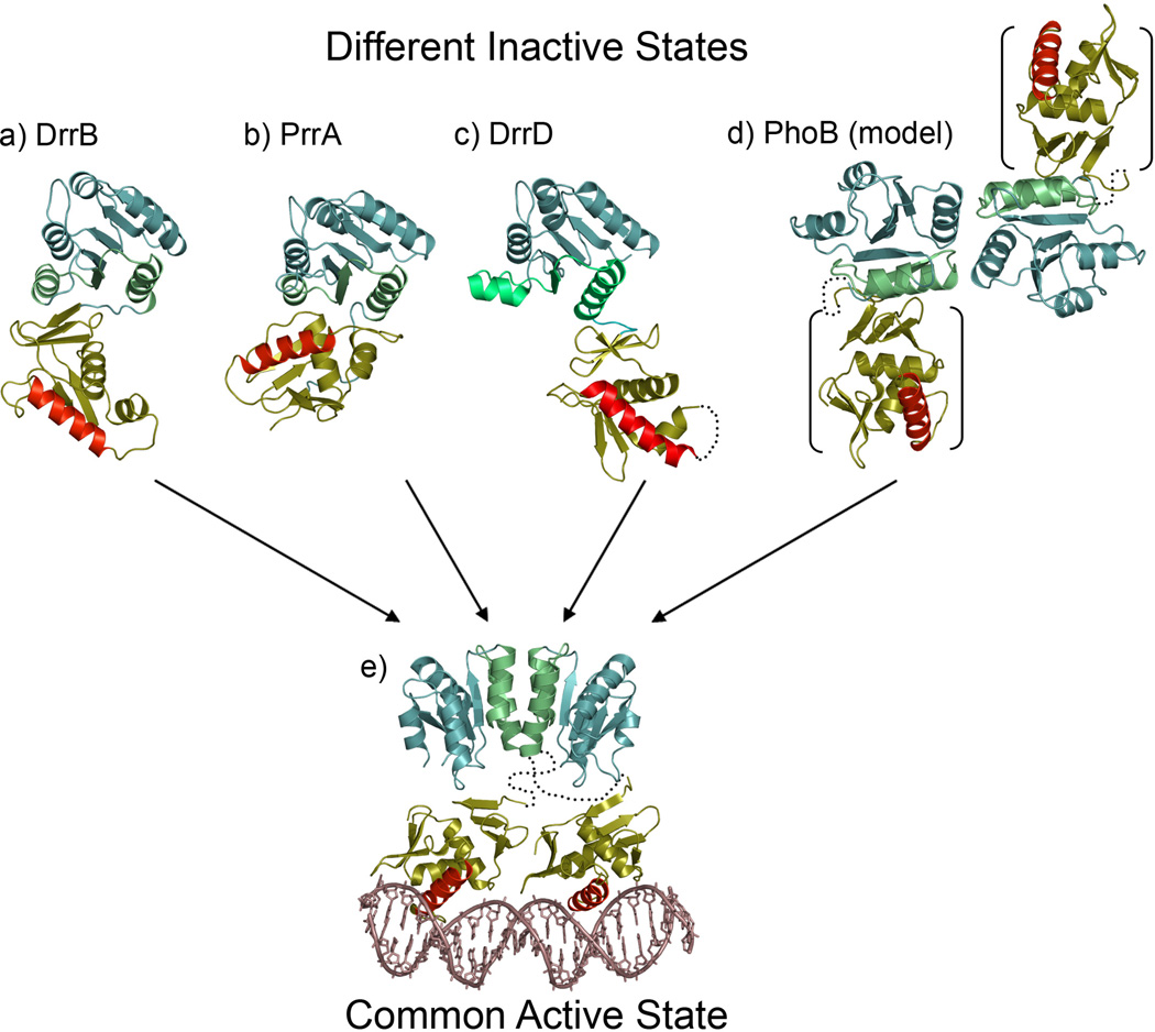
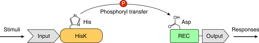
Response regulators (RRs) comprise a major family of signaling proteins in prokaryotes. A modular architecture that consists of a conserved receiver domain and a variable effector domain enables RRs to function as phosphorylation-regulated switches that couple a wide variety of cellular behaviors to environmental cues. Recently, advances have been made in understanding RR functions both at genome-wide and molecular levels. Global techniques have been developed to analyze RR input and output, expanding the scope of characterization of these versatile components. Meanwhile, structural studies have revealed that, despite common structures and mechanisms of function within individual domains, a range of interactions between receiver and effector domains confer great diversity in regulatory strategies, optimizing individual RRs for the specific regulatory needs of different signaling systems.


Figures

Figure 1
Figure 1
Classification of bacterial RRs. RRs are categorized by their effector domains and are further divided into subfamilies based on functions or structures. The distribution of respective types are indicated as percentage values in the set of ~9000 bacterial RRs analyzed by Galperin (http://www.ncbi.nlm.nih.gov/Complete_Genomes/SignalCensus.html) [13].
Figure 2
Figure 2
Conserved features of RR receiver domains. (a) A ribbon diagram of E. coli CheY (1FQW) displays the classic (βα)5 fold of receiver domains with the site of phosphorylation (Asp57) shown in ball-and-stick mode. The α4-β5-α5 signaling face, the region that shows the largest structural perturbations upon phosphorylation, is colored green. (b) A stereo diagram of the active site of CheY (1FWQ) illustrates the roles of highly conserved residues in coordinating the phosphate and divalent metal ion. Due to the lability of the acyl phosphate, structural analyses of active RRs have often been carried out in the presence of the non-covalent phosphoryl analog beryllofluoride [65]. The BeF3 complex (beryllium, orange; fluorides, purple), which coordinates to Asp57, is stabilized by interactions with the side chains of Lys109 and Thr87, and a divalent metal ion (metallic blue). The metal ion (Mn2+ in this structure) is required for catalysis of both phosphotransfer and autodephosphorylation and is positioned by octahedral coordination to the side chains of Asp12 (water-mediated), Asp13, and Asp57, the backbone carbonyl of Asn59, a fluoride of BeF3, and an additional water molecule (green). (c) RRs utilize a common mechanism to couple phosphorylation to surface changes. This mechanism involves the reorientation of two highly conserved residues, a hydroxyl-containing residue (Ser/Thr) and an aromatic residue (Phe/Tyr). The relative orientations of these side chains (Thr87 and Tyr106 in CheY) and that of the phosphorylated aspartate (Asp57) in their inactive (yellow) and active (blue) conformations are shown in ball-and-stick mode following superpositioning of inactive and active CheY structures (2CHE and 1FWQ). In the inactive conformation, the Ser/Thr and Phe/Tyr are oriented away from the active site with the aromatic side chain in a surface exposed position on the α4-β5-α5 face. In the active conformation, both side chains are oriented towards the active site with the Phe/Tyr side chain buried and the hydroxyl group of the Ser/Thr forming a hydrogen bond with a phosphate oxygen (in this structure, a fluoride of BeF3). The view represents a rotation of ~90° about the x-axis relative to the view in (a) and a Cα trace of active CheY with colors as in (a) is shown for reference. In all panels, oxygen atoms are colored red; nitrogen atoms, blue; and carbon atoms, black, unless noted otherwise.
Figure 3
Figure 3
Activation mechanisms of NtrC subfamily RRs. NtrC subfamily members share a similar domain composition of receiver (R), ATPase (C) and DNA-binding (D) domains and are all dependent on the ring assembly of the central ATPase domains (alternating blue and green) to interact with σ54 for transcription regulation. However, despite these similarities, two distinct assembly mechanisms have been discovered within the subfamily so far. In DctD and NtrC1 (top), the central ATPase domain is intrinsically competent for ring assembly but the inactive receiver domain negatively regulates assembly by holding the ATPase (C) domains in a front-to-front dimer, unfavorable for a front-to-back assembly [50]. Phosphorylation or removal of the receiver domain exposes the surface that is buried in the inactive dimer, relieving the inhibition. In NtrC (bottom), the isolated ATPase domain lacks significant ATPase activity. Phosphorylation causes conformational changes that enable the receiver domain from one subunit to be in contact with the ATPase domain of a second subunit, stabilizing the ring structure and promoting ATPase activity [49].
Figure 4
Figure 4
Inactive and active domain arrangements in OmpR/PhoB subfamily members. OmpR/PhoB RRs have different domain orientations in the inactive state, yet all assume a common active state. Structures are available for three inactive full-length multidomain RRs: (a) T. maritima DrrB (1P2F), (b) M. tuberculosis PrrA (1YS6) and (c) T. maritima DrrD (1KGS). Domain arrangements in a fourth RR, (d) E. coli PhoB, can be modeled from structures of the isolated receiver domain dimer (1B00) and the isolated DNA-binding domain (1GXQ). The orientation of the DNA-binding domains (bracketed) relative to the receiver domains in PhoB is unknown, but the short linkers that connect the domains (depicted as dotted lines) restrict placement of the DNA-binding domains to diagonal positions across the receiver domain dimer. Although no structures of active multidomain OmpR/PhoB RRs have been determined, an active state can be readily envisioned (e). The common α4-β5-α5 dimer observed for all active OmpR/PhoB receiver domains paired with a tandem dimer of DNA-binding domains bound to direct repeat half-sites is compatible with only a single active state. The different symmetries of the N- and C-terminal domain dimers preclude a unique intramolecular interface between the domains. Thus flexible linkers (depicted as dotted lines) are proposed to tether the domain dimers, with domain orientations restricted only by the linker length. The depicted model is constructed from structures of the isolated active receiver domain dimer of PhoB (1ZES) and the complex of PhoB DNA-binding domains bound to target DNA (1GXP). Receiver domains are shown in teal with α4-β5-α5 faces highlighted in green; DNA-binding domains are shown in gold with recognition helices highlighted in red.
Box 1, Figure I
Box 1, Figure I
Schematic diagram of a typical two-component system. The conserved His-containing kinase domain of the HPK is shown in orange and the conserved Asp-containing receiver domain of the RR is in green. The variable domains that confer specificity of input (sensing domain of the HPK) and output (effector domain of the RR) to each TCS are shown in grey.

References

    1. Galperin MY. A census of membrane-bound and intracellular signal transduction proteins in bacteria: bacterial IQ, extroverts and introverts. BMC Microbiol. 2005;5:35. - PMC - PubMed
    1. Ulrich LE, Zhulin IB. MiST: a microbial signal transduction database. Nucl. Acids Res. 2007;35:D386–D390. - PMC - PubMed
    1. Ashby MK, Houmard J. Cyanobacterial two-component proteins: structure, diversity, distribution, and evolution. Microbiol. Mol. Biol. Rev. 2006;70:472–509. - PMC - PubMed
    1. Beier D, Gross R. Regulation of bacterial virulence by two-component systems. Curr. Opin. Microbiol. 2006;9:143–152. - PubMed
    1. Kruppa M, Calderone R. Two-component signal transduction in human fungal pathogens. FEMS Yeast Res. 2006;6:149–159. - PubMed

MeSH terms

Substances

LinkOut - more resources