Review
. 2007 Oct;55(2):285-96.
doi: 10.1016/j.brainresrev.2007.02.008. Epub 2007 Mar 12.

Pulvinar contributions to the dorsal and ventral streams of visual processing in primates

Affiliations
Review

Pulvinar contributions to the dorsal and ventral streams of visual processing in primates

Jon H Kaas et al. Brain Res Rev. 2007 Oct.

Abstract

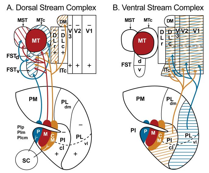
The visual pulvinar is part of the dorsal thalamus, and in primates it is especially well developed. Recently, our understanding of how the visual pulvinar is subdivided into nuclei has greatly improved as a number of histological procedures have revealed marked architectonic differences within the pulvinar complex. At the same time, there have been unparalleled advances in understanding of how visual cortex of primates is subdivided into areas and how these areas interconnect. In addition, considerable evidence supports the view that the hierarchy of interconnected visual areas is divided into two major processing streams, a ventral stream for object vision and a dorsal stream for visually guided actions. In this review, we present evidence that a subset of medial nuclei in the inferior pulvinar function predominantly as a subcortical component of the dorsal stream while the most lateral nucleus of the inferior pulvinar and the adjoining ventrolateral nucleus of the lateral pulvinar are more devoted to the ventral stream of cortical processing. These nuclei provide cortico-pulvinar-cortical interactions that spread information across areas within streams, as well as information relayed from the superior colliculus via inferior pulvinar nuclei to largely dorsal stream areas.


Figures

Figure 1
Figure 1
Surface view of unfolded and flattened cortex of an owl monkey with cortical areas indicated. A view of the intact brain is on the bottom left. Abbreviations: auditory areas: A1, primary auditory; R, rostral area; AT, rostrotemporal area; Aud. B, auditory belt; Aud. P.B., auditory parabelt. Somatosensory areas: 3a, 3b, 1 and 2 of Brodmann (3a = primary somatosensory area, S-1), S2, second area; PV, parietal ventral area; PR, parietal rostral area; VS, parietal ventral area. Motor areas: rostral, r, and caudal, c, divisions of primary motor cortex, M1; PMD, dorsal premotor cortex; PMV, ventral premotor cortex; SMAd, dorsal supplementary motor area; SMAm, medial supplementary motor area; E-SMA, eye-supplementary motor area; CMr, rostral cingulate motor area; CMc, caudal cingulate motor area; FEF, frontal eye field; FV, frontal visual area. Visual areas: V1, V2, and V3, first, second and third visual areas; M, medial visual area; DM, dorsomedial visual area; DLr and DLc, rostral and caudal dorsolateral visual areas; ITc, ITr, ITm, and ITp, caudal, rostral, medial, and polar divisions of inferior temporal (IT) cortex; MT, the middle temporal visual area; MTc, the middle temporal crescent; MST, the middle superior temporal area; FSTv and FSTd, ventral and dorsal areas of the fundus of the superior temporal sulcus (FST); VPP, ventral posterior parietal region; PP, posterior parietal cortex. Also, cc, corpus callosum.
Figure 2
Figure 2
Subdivisions and connections of the pulvinar complex in macaque monkeys. The outline is from a coronal section through the complex with lateral to the right. The medial pulvinar (PM), with medial (m) and lateral (l) divisions, and the dorsomedial nucleus of the lateral pulvinar (PLdm) have connections with dorsolateral (DL) prefrontal cortex and other parietal, frontal, and temporal areas, and are not considered further here. The ventrolateral nucleus of the lateral pulvinar (Plvl) and the central lateral nucleus of the inferior pulvinar (PIcl) contain large retinotopic representations of the contralateral visual hemifield (+, upper field; - lower field), and interconnect with areas more devoted to the ventral stream of visual processing, while the posterior (p), medial (m), and central medial (cm) nuclei of the inferior pulvinar (PIp, PIm, and PIcm) interconnect with areas of the dorsal stream. See Gutierrez et al. (2000) for a detailed account of the connections of PM and PLdm. See Stepniewska (2003) for review.
Figure 3
Figure 3
The retinotopy of nuclei of the inferior and lateral pulvinar. Squares mark the representation of the zero horizontal meridian, while circles mark the vertical meridian. The locations of -10° and +15° on PLvl mark approximate locations representing 10° in the lower field and 15° in the upper field, respectively. (+, upper field; - lower field) Coronal brain section with lateral to the right. Based on unpublished studies of Lyon and Kaas and Adams et al. (2000).
Figure 4
Figure 4
The inferior pulvinar of a squirrel monkey in a coronal section (lateral to the right) processed for acetylcholinesterase (AchE). The posterior (PIp), medial (PIm), central medial (PIcm) and central lateral nuclei (PIcl) of the inferior pulvinar are identified. A lighter lateral zone in PIcl may correspond to a “shell” (s) division of the inferior pulvinar proposed by Gutierrez et al. (1995). The dorsal lateral geniculate nucleus (LGN) is on the right, and the lateral pulvinar (PL) above. Br. CS, brachium of the superior colliculus. Based on Stepniewska and Kaas, 1997.
Figure 5
Figure 5
Histological subdivisions of the inferior pulvinar in New World owl and squirrel monkeys and Old World macaque monkeys. The subdivisions are based on the results of a number of studies including Nissl and myelin stains, and reactions for acetylcholinesterase (AchE), Calbindin (Cb), cytochrome oxidase (CO) and the antigen for the antibody Cat-301. The figure has been modified from Stepniewska and Kaas (1979).
Figure 6
Figure 6
Evidence for topographic patterns of connections of primary visual cortex (V1) with the ventral lateral nucleus of the lateral pulvinar (PL) and the central lateral nucleus of the inferior pulvinar (PIcl) of a macaque monkey. Injection of different tracers (see Lyon and Kaas, 2002c for details) were placed in locations above (-10°) and below (+15°) the calcarine fissure in V1 that labeled neurons in -10° and +15° locations in PL and PIcl. The box on the upper right shows the locations of the two injection sites on a caudodorsal view of the right cerebral hemisphere. CC, corpus callosum; Cb, cerebellum; Calc, calcarine sulcus; LuS, lunate sulcus; STS, superior temporal sulcus; IPS, intraparietal sulcus; LS, lateral sulcus. The box on the lower right shows the retinotopic locations of the injections in a schematic of the contralateral visual hemifield. On the left, nuclei of the pulvinar complex are outlined on an anterior to posterior numbered series of coronal brain sections. Dots indicate the locations of labeled cells. Connections as in previous figures.
Figure 7
Figure 7
Thalamic neurons labeled by an injection in V2, MST, or primary auditory cortex (A1) of a marmoset. Thalamic neurons labeled by each injection are indicated by a different symbol (V2, filled squares; MST, open squares, A1, triangles. Conventions are as in previous figures.
Figure 8
Figure 8
Thalamic neurons labeled by an injection in MT (green triangles), the rostral DM border (orange squares), the rostral border of MST (blue squares), or MST (open red squares). Conventions as in previous figures.
Figure 9
Figure 9
Thalamic connections labeled by injections in FSTd and MST. Labeled neurons are largely in PIm.
Figure 10
Figure 10
Nuclei of the pulvinar related to visual areas of the dorsal and ventral streams. A. Arrows indicate connections between nuclei of the inferior pulvinar and cortical areas of the dorsal stream. Note also, the superior colliculus inputs to posterior (p) and central medial (cm) nuclei of the inferior pulvinar (PI). B. Arrows indicate connections between visual areas of the ventral stream and the central lateral nucleus of the inferior pulvinar (PIcl) and the ventrolateral nucleus of the lateral pulvinar (PL).

References

    1. Adams AD, Hof PR, Gattass R, Webster MJ, Ungerleider LG. Visual cortical projections and chemoarchitecture of macaque monkey pulvinar. J. Comp. Neurol. 2000;419:377–393. - PubMed
    1. Allman JM, Kaas JH. A representation of a visual field in the caudal third of the middle temporal gyrus of the owl monkey (Aotus trivirgatus) Brain Res. 1971;31:85–105. - PubMed
    1. Allman JM, Kaas JH. A crescent-shaped cortical visual area surrounding the middle temporal area (MT) in the owl monkey (Aotus trivirgatus) Brain Res. 1974;81:199–213. - PubMed
    1. Allman JM, Kaas JH. The dorsomedial cortical visual area: A third tier area in the occipital lobe of the owl monkey (Aotus trivirgatus) Brain Res. 1975;100:473–487. - PubMed
    1. Baizer JS, Desimone R, Ungerleider LG. Comparison of subcortical connections of inferior temporal and posterior parietal cortex in monkeys. Vis. Neurosci. 1993;10:59–72. - PubMed

LinkOut - more resources