

The effects of brain tissue decomposition on diffusion tensor imaging and tractography
- PMID: 17433879
- PMCID: PMC4039353
- DOI: 10.1016/j.neuroimage.2007.02.039
The effects of brain tissue decomposition on diffusion tensor imaging and tractography
Abstract
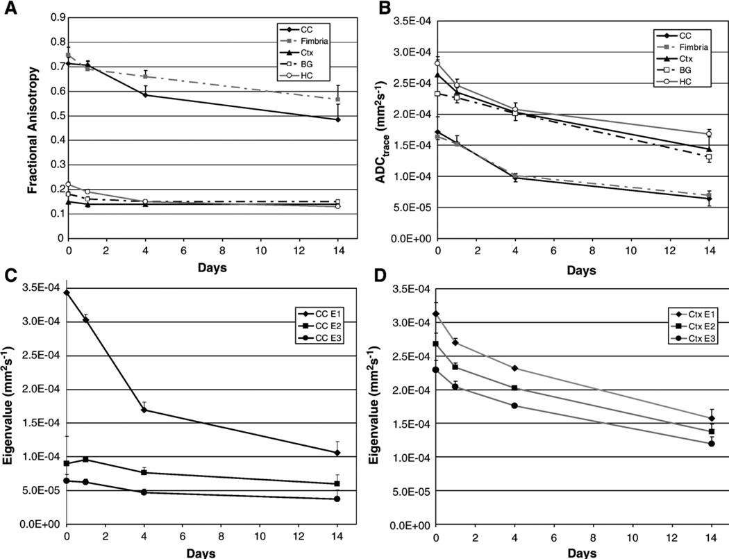
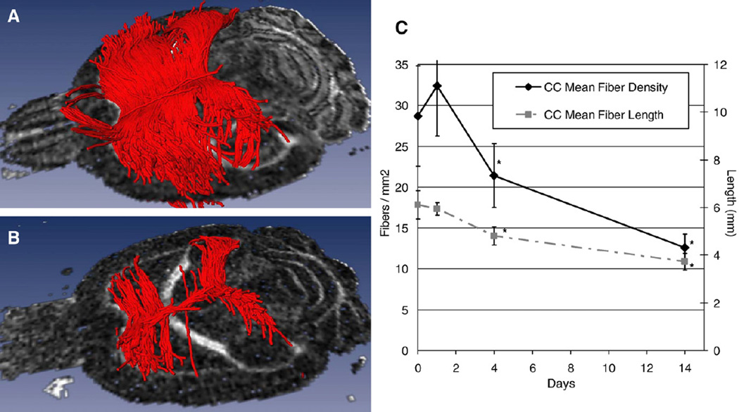
There have been numerous high resolution diffusion tensor imaging studies in fixed animal brains, but relatively few studies in human brains. While animal tissues are generally fixed pre-mortem or directly postmortem, this is not possible for human tissue, therefore there is always some delay between death and tissue fixation. The elapsed time between death and tissue fixation, the postmortem interval (PMI), will most likely adversely affect the tissue's diffusion properties. We studied the effects of PMI on the diffusion properties of rodent brain. Eight mice were euthanized and the brains (kept in the skull) were placed in formalin at PMIs of 0, 1, 4 and 14 days. Post fixation they were placed in a solution of GdDTPA and phosphate buffered saline. Brains were scanned with a 3D EPI DTI sequence at 4.7T. DTI data were processed to generate apparent diffusion coefficient (ADC) and fractional anisotropy (FA) maps. DTI tractography was also performed. The temporal changes in regional ADC and FA values were analyzed statistically using a one-way ANOVA, followed by individual Student's T-tests. Regional FA and ADC of gray and white matter decreased significantly with time (p<0.05). DTI tractography showed a decrease in the number and coherence of reconstructed fiber pathways between PMIs 0 and 14. Elapsed time between death and tissue fixation has a major effect upon the brain's diffusion properties and should be born in mind when interpreting fixed brain DTI.
Figures
References
-
- Bar W, Kratzer A, Machler M, Schmid W. Postmortem stability of DNA. Forensic Sci. Int. 1988;39(1):59–70. - PubMed
-
- D'Arceuil H, Grant E, de Crespigny AJ. High resolution ex vivo Diffusion Tensor Imaging (DTI) and tractography in the developing rabbit brain; Radiological Society of North America (RSNA) Annual Meeting; Chicago, IL. 2005. SSQ15–01.
-
- D'Arceuil HE, Westmoreland S, de Crespigny AJ. An approach to high resolution diffusion tensor imaging in fixed primate brain. NeuroImage. 2007;35(2):553–565. - PubMed
-
- Guilfoyle DN, Helpern JA, Lim KO. Diffusion tensor imaging in fixed brain tissue at 7.0 T. NMR Biomed. 2003;16(2):77–81. - PubMed
-
- Huang H, Zhang J, Wakana S, Zhang W, Ren T, Richards LJ, Yarowsky P, Donohue P, Graham E, van Zijl PC, Mori S. White and gray matter development in human fetal, newborn and pediatric brains. NeuroImage. 2006;33(1):27–38. - PubMed
Publication types
MeSH terms
Grants and funding
LinkOut - more resources
Full Text Sources
Other Literature Sources
