. 2007 Aug 1;364(2):383-94.
doi: 10.1016/j.virol.2007.03.017. Epub 2007 Apr 16.

ICAM-3 influences human immunodeficiency virus type 1 replication in CD4(+) T cells independent of DC-SIGN-mediated transmission

Affiliations

ICAM-3 influences human immunodeficiency virus type 1 replication in CD4(+) T cells independent of DC-SIGN-mediated transmission

Julia E Biggins et al. Virology. .

Abstract

We investigated the role of ICAM-3 in DC-SIGN-mediated human immunodeficiency virus (HIV) infection of CD4(+) T cells. Our results demonstrate that ICAM-3 does not appear to play a role in DC-SIGN-mediated infection of CD4(+) T cells as virus is transmitted equally to ICAM-3(+) or ICAM-3(-) Jurkat T cells. However, HIV-1 replication is enhanced in ICAM-3(-) cells, suggesting that ICAM-3 may limit HIV-1 replication. Similar results were obtained when SIV replication was examined in ICAM-3(+) and ICAM-3(-) CEMx174 cells. Furthermore, while ICAM-3 has been proposed to play a co-stimulatory role in T cell activation, DC-SIGN expression on antigen presenting cells did not enhance antigen-dependent activation of T cells. Together, these data indicate that while ICAM-3 may influence HIV-1 replication, it does so independent of DC-SIGN-mediated virus transmission or activation of CD4(+) T cells.


Figures

Fig. 1
Fig. 1. Selection and infection of ICAM-3 negative Jurkat cell line
(A) Phenotype of wildtype and ICAM-3 Jurkat cells. The wildtype Jurkat cells (ICAM-3+) and mutant (ICAM-3) cells were examined for surface expression of ICAM-3, CD3, CD28, CD4, and CXCR4 (closed curves). Isotype control antibodies are represented by open curves. The mean fluorescence intensity (MFI) of the stained cells is indicated on each histogram. (B) HIV-1 infection of wildtype and ICAM-3 negative Jurkat cells. Jurkat cells were incubated with an HIV-luciferase virus pseudotyped with a CXCR4-tropic HIV-1 envelope or left untreated for 3 hours. On the third day post-infection, cells were harvested and virus production was analyzed by measuring luciferase activity in the infected cell lysates. Mock treated cells were used for background controls. Relative luciferase activity ± standard error of the mean (SEM) is shown.
Fig. 2
Fig. 2. DC-SIGN mediated transmission of HIV-1 to ICAM-3 positive and negative Jurkat cells
(A) Pig-tailed macaque DC-SIGN expressing Raji cells were created via retroviral transduction. DC-SIGN expression was confirmed through FACS analysis by using an anti-DC-SIGN monoclonal antibody (filled curves) or isotype control antibody (open curves). (B) Capture and transmission of HIV from DC-SIGN+ Raji cells to ICAM-3 Jurkat cells. DC-SIGN+ Raji cells were incubated with a pseudotyped HIV-luciferase virus for 2 hours, washed and then co-cultured with target cells. On the third day post infection virus production was analyzed by measuring luciferase activity in target cells. (C) Capture and transmission of HIV-1 from DC-SIGN+ Raji cells to ICAM-3 Jurkat cells in the presence of superantigen. DC-SIGN mediated virus capture and transfer assay was performed as described above. Co-cultures were maintained in the presence or absence of staphylococcal enterotoxin D (SED). (D) Capture and transmission of HIV-1 from primary human dendritic cells to ICAM-3 Jurkat cell line. A DC-SIGN mediated virus capture and transfer assay was performed as described above except that human monocyte derived dendritic cells were substituted for DC-SIGN+ Raji cells. All of the experiments were performed in triplicate and the data are representative of at least two independent experiments. Mock treated cells were used for background direct virus infection. Relative luciferase activity ± SEM is shown.
Fig. 3
Fig. 3. HIV-1 replication is enhanced in the absence of ICAM-3
ICAM-3 or ICAM-3+ Jurkat cells were infected with HIV-1NL4-3 at a low multiplicity of infection (MOI). At various times during a two-week period post-infection supernatants were collected and virus production was measured by p24 antigen capture ELISA. Experiments were performed in triplicate, and the data are representative of three independent experiments. The average levels of p24gag ± SEM are shown.
Fig. 4
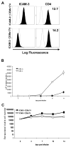
Fig. 4. Selection and infection of ICAM-3 negative CEMx174 cells
(A) Phenotype of wildtype and ICAM-3 CEMx174 cells. The wildtype CEMx174 cells (ICAM-3+) and mutant (ICAM-3) cells were examined for surface expression of ICAM-3 and CD4 (closed curves). Isotype control antibodies are represented by open curves. The mean fluorescence intensity (MFI) of the stained cells is indicated on each histogram. (B) SIV infection of wildtype and ICAM-3 CEMx174 cells. (C) Extrapolated viable cell number of wild type and ICAM-3 CEMx174 cells. CEMx174 cells were infected with SIVmne170 at a low MOI. At various times during a two-week time period post-infection supernatants were collected and virus production was measured using an SIV p27 antigen capture ELISA. The average levels of p27gag ± SEM are shown.
Fig. 5
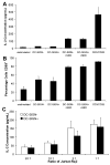
Fig. 5. DC-SIGN does not modulate T cell activation
(A) IL-2 concentration in supernatants after activation. (B) CD69 surface expression on Jurkat cells. Jurkat cells were either cultured alone and unstimulated, co-cultured with either DC-SIGN+ or DC-SIGN Raji cells with or without Staphylococcal enterotoxin D (SED), or cultured alone and treated with anti-CD3 and anti-CD28 antibodies. The average IL-2 levels ±SEM are shown. (C) Activation of Jurkat cells in the presence of different amounts of DC-SIGN+ Raji cells. Jurkat cells were co-cultured with either DC-SIGN+ or DC-SIGN Raji cells and SED at the indicated ratios. After 24 hours, culture supernatants were examined for IL-2 concentration by ELISA. The average IL-2 levels ±SEM are shown.
Fig. 6
Fig. 6. Soluble DC-SIGN does not co-stimulate T cells
(A) Expression of sDC-SIGN. Conditioned supernatants from S2 cells stably expressing sDC-SIGN were collected and subjected to SDS-12% polyacrlyamide gel electrophoresis (PAGE). A western blot was performed and the His-tagged protein was detected by enhanced chemiluminescence. Cells were either left untreated (−) or induced to express DC-SIGN with copper sulfate (+). (B) sDC-SIGN inhibition of virion capture. DC-SIGN expressing 293T cells were incubated with indicated inhibitors for 20 minutes. The cells were then incubated with SIVmne170 for 2 hours. Cells were washed, lysed, and the amount of virus remaining bound to the cell surface was determined by antigen capture ELISA. Mock treated cells were used for background controls. Virus captured in the presence of each inhibitor is shown relative to untreated cells after correcting for background binding ± SEM. (C) Co-stimulation Assay. Jurkat cells were added to wells coated with indicated amounts of anti-CD3 antibody alone (■), anti-CD3 antibody + 1 μg sDC-SIGN (▒), coated with anti-CD3 antibody only and 1 μg sDC-SIGN added to culture medium (▧ ) or coated with anti-CD3 + 1 μg anti-CD28 antibodies (□). After 24 hours culture supernatants were examined for IL-2 concentration by ELISA. (D) ICAM-3 Activation. Jurkat cells were added to wells left untreated or coated with 1 μg/well each of anti-CD3 antibody, anti-CD3 antibody and sDC-SIGN, anti-CD3 antibody only with sDC-SIGN added to culture supernatant, anti-CD3 and anti-ICAM-3 (ICR1.1) antibodies, or anti-CD3 and anti-CD28 antibodies. After 24 hours, IL-2 concentration was measured in supernatants by ELISA. The average IL-2 levels ±SEM are shown. Student’s t-test was performed: p< .001.
Fig. 7
Fig. 7. IL-2 production is sustained despite loss of ICAM-3-DC-SIGN interaction
Wildtype or ICAM-3 Jurkat cells were cocultured with either wildtype or DC-SIGN+ Raji cells in the presence or absence of Staphylococcal enterotoxin D (SED). Alternately, Jurkat cells cultured alone were treated with anti-CD3 and anti-CD28 antibodies or left untreated. After 24 hours, IL-2 supernatant levels were analyzed by ELISA. The average IL-2 levels ±SEM are shown.

Similar articles

Cited by

References

    1. Arrighi JF, Pion M, Garcia E, Escola JM, van Kooyk Y, Geijtenbeek TB, Piguet V. DC-SIGN-mediated Infectious Synapse Formation Enhances X4 HIV-1 Transmission from Dendritic Cells to T Cells. J Exp Med. 2004;200:1279–1288. - PMC - PubMed
    1. Barat C, Gervais P, Tremblay MJ. Engagement of ICAM-3 Provides a Costimulatory Signal for Human Immunodeficiency Virus Type 1 Replication in both Activated and Quiescent CD4+ T Lymphocytes: Implications for Virus Pathogenesis. J Virol. 2004;78:6692–6697. - PMC - PubMed
    1. Baribaud F, Pohlmann S, Sparwasser T, Kimata MTY, Choi YK, Haggarty BS, Ahmad N, Macfarlan T, Edwards TG, Leslie GJ, Arnason J, Reinhart TA, Kimata JT, Littman DR, Hoxie JA, Doms RW. Functional and Antigenic Characterization of Human, Rhesus Macaque, Pigtailed Macaque, and Murine DC-SIGN. J Virol. 2001;75:10281–10289. - PMC - PubMed
    1. Berney SM, Schaan T, Alexander JS, Peterman G, Hoffman PA, Wolf RE, van der Heyde H, Atkinson TP. ICAM-3 (CD50) cross-linking augments signaling in CD3-activated peripheral human T lymphocytes. J Leukoc Biol. 1999;65:867–874. - PubMed
    1. Biggins JE, Yu Kimata MT, Kimata JT. Domains of macaque DC-SIGN essential for capture and transfer of simian immunodeficiency virus. Virology. 2004;324:194–203. - PubMed

Publication types

MeSH terms