

Molecular imaging with targeted perfluorocarbon nanoparticles: quantification of the concentration dependence of contrast enhancement for binding to sparse cellular epitopes
- PMID: 17434667
- PMCID: PMC1978071
- DOI: 10.1016/j.ultrasmedbio.2006.12.007
Molecular imaging with targeted perfluorocarbon nanoparticles: quantification of the concentration dependence of contrast enhancement for binding to sparse cellular epitopes
Abstract
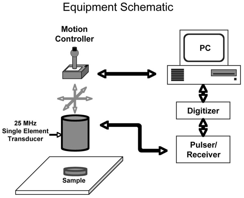
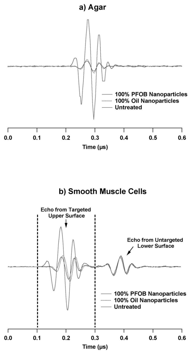
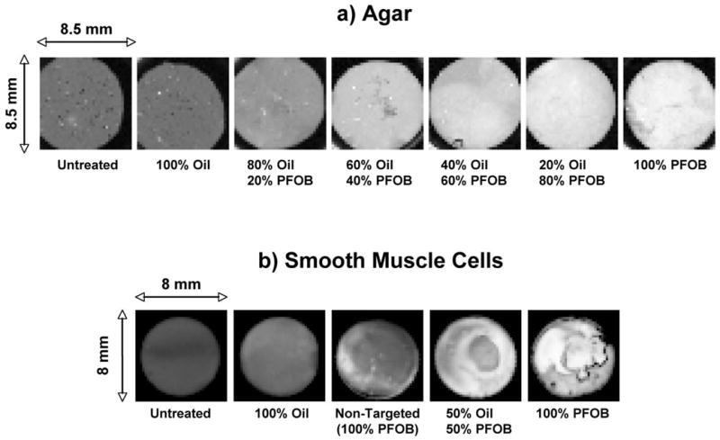
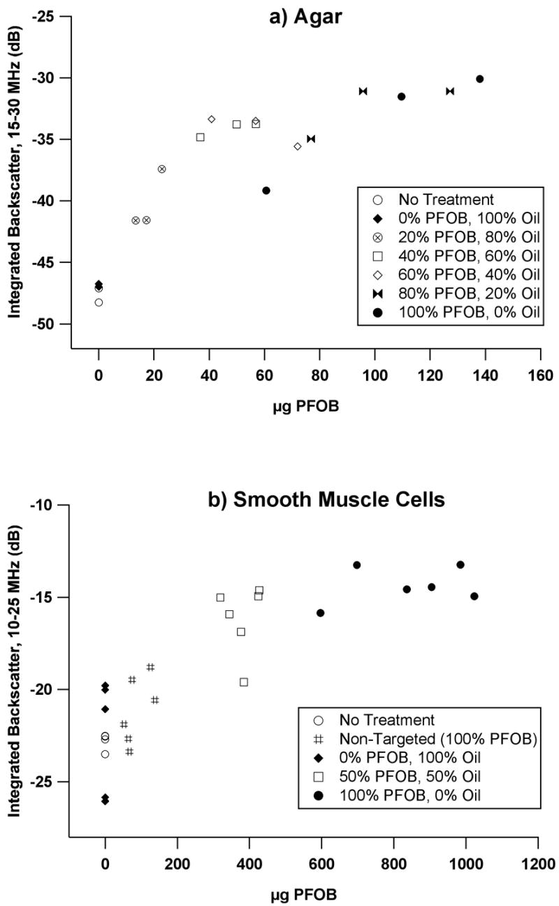
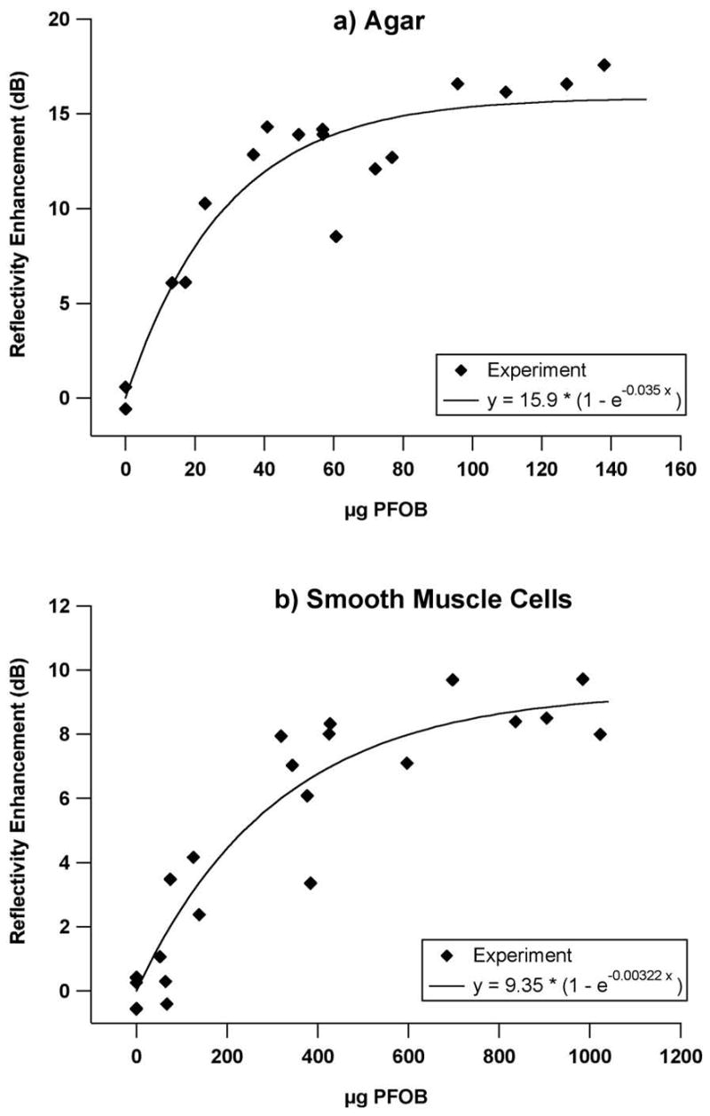
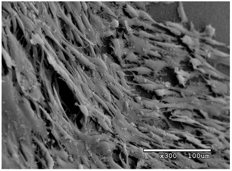
Targeted, liquid perfluorocarbon nanoparticles are effective agents for acoustic contrast enhancement of abundant cellular epitopes (e.g., fibrin in thrombi) and for lower prevalence binding sites, such as integrins associated with tumor neovasculature. In this study, we sought to delineate the quantitative relationship between the extent of contrast enhancement of targeted surfaces and the density (and concentration) of bound perfluorocarbon (PFC) nanoparticles. Two dramatically different substrates were utilized for targeting. In one set of experiments, the surfaces of smooth, flat, avidin-coated agar disks were exposed to biotinylated nanoparticles to yield a thin layer of targeted contrast. For the second set of measurements, we targeted PFC nanoparticles applied in thicker layers to cultured smooth muscle cells expressing the transmembrane glycoprotein "tissue factor" at the cell surface. An acoustic microscope was used to characterize reflectivity for all samples as a function of bound PFC (determined via gas chromatography). We utilized a formulation of low-scattering nanoparticles having oil-based cores to compete against high-scattering PFC nanoparticles for binding, to elucidate the dependence of contrast enhancement on PFC concentration. The relationship between reflectivity enhancement and bound PFC content varied in a curvilinear fashion and exhibited an apparent asymptote (approximately 16 dB and 9 dB enhancement for agar and cell samples, respectively) at the maximum concentrations (approximately 150 microg and approximately 1000 microg PFOB for agar and cell samples, respectively). Samples targeted with only oil-based nanoparticles exhibited mean backscatter values that were nearly identical to untreated samples (<1 dB difference), confirming the oil particles' low-scattering behavior. The results of this study indicate that substantial contrast enhancement with liquid perfluorocarbon nanoparticles can be realized even in cases of partial surface coverage (as might be encountered when targeting sparsely populated epitopes) or when targeting surfaces with locally irregular topography. Furthermore, it may be possible to assess the quantity of bound cellular epitopes through acoustic means.
Figures
References
-
- Alkan-Onyuksel H, Demos SM, Lanza GM, et al. Development of inherently echogenic liposomes as an ultrasonic contrast agent. J Pharm Sci. 1996;85:486–90. - PubMed
-
- Chamley-Campbell J, Campbell GR, Ross R. The smooth muscle cell in culture. Physiol Rev. 1979;59:1–61. - PubMed
-
- Couture O, Bevan PD, Cherin E, et al. A model for reflectivity enhancement due to surface bound submicrometer particles. Ultrasound Med Biol. 2006;32:1247–55. - PubMed
-
- Hall CS, Marsh JN, Scott MJ, et al. Temperature dependence of ultrasonic enhancement with a site-targeted contrast agent. Journal of the Acoustical Society of America. 2001;110:1677–84. - PubMed
-
- Hughes MS, Marsh JN, Zhang H, et al. Characterization of digital waveforms using thermodynamic analogs: detection of contrast-targeted tissue in vivo. IEEE Trans Ultrason Ferroelectr Freq Control. 2006;53:1609–16. - PubMed
Publication types
MeSH terms
Substances
Grants and funding
LinkOut - more resources
Full Text Sources
Other Literature Sources
Miscellaneous
