Review
. 2007 Jul;92(4):635-40.
doi: 10.1113/expphysiol.2006.036376. Epub 2007 Apr 13.

Signalling within the neurovascular unit in the mammalian retina

Affiliations
Review

Signalling within the neurovascular unit in the mammalian retina

Monica R Metea et al. Exp Physiol. 2007 Jul.

Abstract

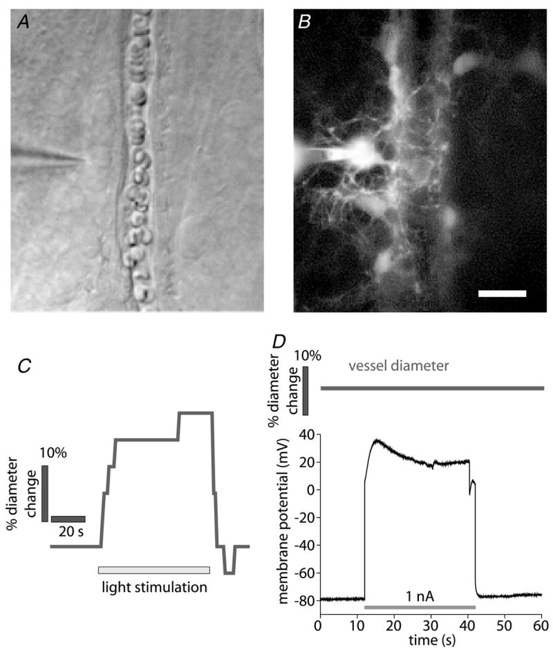
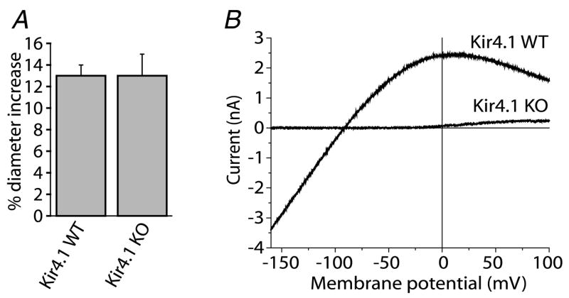
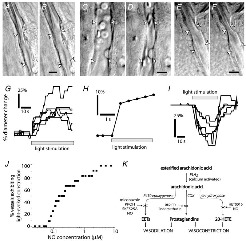
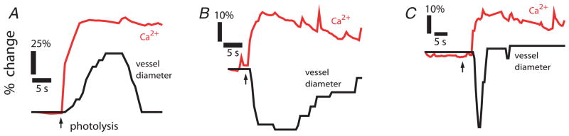
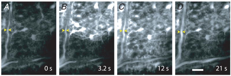
Neuronal activity in the central nervous system evokes localized changes in blood flow, a response termed neurovascular coupling or functional hyperaemia. Modern functional imaging methods, such as functional magnetic resonance imaging (fMRI), measure signals related to functional hyperaemia in order to determine localization of brain function and to diagnose disease. The cellular mechanisms that underlie functional hyperaemia, however, are not well understood. Glial cells have been hypothesized to be intermediaries between neurons and blood vessels in the control of neurovascular coupling, owing to their ability to release vasoactive factors in response to neuronal activity. Using an in vitro preparation of the isolated, intact rodent retina, we have investigated two likely mechanisms of glial control of the vasculature: glial K(+) siphoning and glial induction of vasoactive arachidonic acid metabolites. Potassium siphoning is a process by which a K(+) current flowing through glial cells transfers K(+) released from active neurons to blood vessels. Since slight increases in extracellular K(+) can cause vasodilatation, this mechanism was hypothesized to contribute to neurovascular coupling. Our data, however, suggest that glial K(+) siphoning does not contribute significantly to neurovascular coupling in the retina. Instead, we suggest that glial cells mediate neurovascular coupling by inducing the production of two types of arachidonic acid metabolites, epoxyeicosatrienoic acids (EETs) and 20-hydroxyeicosatetraenoic acid (20-HETE), which dilate and constrict vessels, respectively. We show that both light flashes and direct glial stimulation produce vasodilatation or vasoconstriction mediated by EETs and 20-HETE, respectively. Further, we show that the type of vasomotor response observed (dilatation or constriction) depends on retinal levels of nitric oxide. Our data also demonstrate that glial cells are necessary intermediaries for signalling from neurons to blood vessels, since functional hyperaemia does not occur when neuron-to-glia communication is interrupted. These results indicate that glial cells play an important role in mediating functional hyperaemia and suggest that the regulation of blood flow may involve both vasodilating and vasoconstricting components.


Figures

Figure 1
Figure 1. Light stimulation, but not glial cell depolarization, evokes vasodilatation
A and B, micrographs showing a whole cell-patched astrocyte contacting an arteriole. The patch pipette is seen at the left. A, infrared-differential interference contrast (IR-DIC) image. B, fluorescence image showing the Lucifer Yellow-filled astrocyte contacting the arteriole. Additional astrocytes coupled to the patched cell are also seen. Scale bar for A and B represents 10 μm. C, time course of vessel diameter change. Flickering light stimulation evokes vasodilatation, demonstrating neurovascular coupling in the retina. D, the astrocyte shown in A and B is depolarized by injection of 1 nA current. Arteriole diameter does not change during the depolarization, indicating that K+ siphoning is not sufficient to initiate vasodilatation. In a series of experiments, glial cells were depolarized to membrane potentials ranging from −40 to +120 mV, sufficient to produce K+ siphoning in many glial cells coupled together. Each cell was depolarized multiple times. No change in arteriole diameter was observed during glial depolarization.
Figure 2
Figure 2. Light-evoked vasodilatation is not reduced in Kir4.1 knockout (KO) mice despite the absence of K+ currents in retinal glial cells
A, flickering light stimulation evokes vasodilatation of similar amplitude in both wild-type (WT) and Kir4.1 knockout mice. B, Ba2+-sensitive current–voltage relations of Müller cells from Kir4.1 WT and KO mice. Ba2+-sensitive inward current is completely absent in the KO cell.
Figure 3
Figure 3. Light stimulation evokes vasodilatation and vasoconstriction, an in vitro model of neuro-vascular coupling in the retina
A–F, IR-DIC images of arterioles at the vitreal surface of the retina. Shown are vessels before (A) and during light-evoked vasodilatation (B); before (C) and during light-evoked constriction (D); and before (E) and during light-evoked sphincter-like constriction (F). Scale bars represent 10 μm. G, time course of light-evoked vasodilatation in 6 trials. H, time course of vasodilatation can be very fast (latency < 500 ms). I, time course of vasoconstriction in 5 trials. J, the concentration of NO determines the type of vascular response to light stimulation. Raising NO levels increases the percentage of vessels which constrict to light. Below 70 nM NO, light stimulation evokes vasodilatation in all vessels (n = 12). As NO is raised, a greater percentage of vessels constrict. K, schematic diagram of vasoactive metabolites of arachidonic acid. See Metea & Newman (2006) for details.
Figure 4
Figure 4. Activation of glial cells produces the same types of vascular responses as light stimulation
Glial cells were stimulated by UV photolysis of caged-Ca2+ within the cells. Photolysis evoked propagated Ca2+ increases in networks of glial cells apposed to blood vessels. These Ca2+ increases were correlated with vessel dilatation (A), constriction (B) or sphincter-like constriction (C). These glial-evoked vasomotor responses occurred even when transmitter release from neurones was blocked by tetanus toxin.
Figure 5
Figure 5. Propagated glial Ca2+ waves evoke vasodilatation in distant arterioles
A–D, a Ca2+ wave produced by ejection of ATP propagates through glial cells at the surface of the retina. The Ca2+ wave dilates an arteriole when it reaches the vessel. The site of focal ATP ejection, used to initiate the Ca2+ wave, was just beyond the upper right corner of the images. Scale bar represents 20 μm.

References

    1. Badeer HS. Hemodynamics for medical students. Adv Physiol Educ. 2001;25:44–52. - PubMed
    1. Brew H, Gray PTA, Mobbs P, Attwell D. Endfeet of retinal glial cells have higher densities of ion channels that mediate K+ buffering. Nature. 1986;324:466–468. - PubMed
    1. Butt AM, Kamada T. Inwardly rectifying potassium channels (Kir) in central nervous system glia: a special role for Kir4.1 in glial functions. J Cell Mol Med. 2006;10:33–44. - PMC - PubMed
    1. Chrissobolis S, Sobey CG. Inwardly rectifying potassium channels in the regulation of vascular tone. Curr Drug Targets. 2003;4:281–289. - PubMed
    1. Chrissobolis S, Ziogas J, Chu Y, Faraci FM, Sobey CG. Role of inwardly rectifying K+ channels in K+-induced cerebral vasodilatation in vivo. Am J Physiol Heart Circ Physiol. 2000;279:H2704–H2712. - PubMed

Publication types

Substances