. 2007 Jul 1;93(1):176-82.
doi: 10.1529/biophysj.106.099929. Epub 2007 Apr 13.

DNA compaction by the nuclear factor-Y

Affiliations

DNA compaction by the nuclear factor-Y

Rosalinda F Guerra et al. Biophys J. .

Abstract

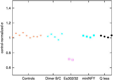
The nuclear factor-Y (NF-Y), a trimeric, CCAAT-binding transcriptional activator with histone-like subunits, was until recently considered a prototypical promoter transcription factor. However, recent in vivo chromatin immunoprecipitation assays associated with microarray methodologies (chromatin immunoprecipitation on chip experiments) have indicated that a large portion of target sites (40%-50%) are located outside of core promoters. We applied the tethered particle motion technique to the major histocompatibility complex class II enhancer-promoter region to characterize i), the progressive compaction of DNA due to increasing concentrations of NF-Y, ii), the role of specific subunits and domains of NF-Y in the process, and iii), the interplay between NF-Y and the regulatory factor-X, which cooperatively binds to the X-box adjacent to the CCAAT box. Our study shows that NF-Y has histone-like activity, since it binds DNA nonspecifically with high affinity to compact it. This activity, which depends on the presence of all trimer subunits and of their glutamine-rich domains, seems to be attenuated by the transcriptional cofactor regulatory factor-X. Most importantly NF-Y-induced DNA compaction may facilitate promoter-enhancer interactions, which are known to be critical for expression regulation.


Figures

FIGURE 1
FIGURE 1
Schematic representation of the fragments of DNA and the proteins used. (A) Ea represents the fragment of DNA where the Y elements are the two CCAAT boxes recognized by NF-Y and the X elements are specific sites for RFX binding, and Ea302/32 represents a similar fragment containing mutated CCAAT boxes, which prevent NF-Y binding. (B) Schematic representation of the three subunits constituting the heterotrimeric complex of NF-Y. HFM stands for histone fold motif; YA9, YB4, and YC5 are core regions highly conserved, and Q indicates a Q-rich domain (bottom).
FIGURE 2
FIGURE 2
Schematic representation of the TPM experimental setup. (Left) a submicron-size bead is tethered to the glass surface of a flow microchamber by a single DNA molecule. Addition of NF-Y induces a change in the effective length of the tethering DNA, causing a decrease in the average Brownian motion of the microsphere measured by σ. This difference is visualized as the amplitude of the Brownian motion of the bead as a function of time.
FIGURE 3
FIGURE 3
Effect of the concentration increase of NF-Y on Ea. (A) The root mean-square displacement (σ) of the microsphere is plotted versus the time of acquisition. Each color represents the trace relative to a single microsphere bound to DNA. Yellow is the control in the absence of NF-Y; turquoise, blue, and purple are in the presence of 1.2, 1.8, and 3.6 nM NF-Y respectively. (B) The average root mean-square displacement is plotted for individual microspheres (A, B, C, …). Each point represents the average value of the Brownian motion of one microsphere tethered by a single DNA molecule. The same letter represents the same microsphere in conditions of increasing concentrations of NF-Y. The stepped line at the bottom of (B) indicates different concentrations of NF-Y. The standard deviation is often too small to be visible.
FIGURE 4
FIGURE 4
Effect of mutant NF-Y proteins and the CCAAT box on compaction. Here, the y axis reports the root mean-square displacement, σ, which is normalized to the value in the absence of NF-Y. The red dots refer to DNA in the absence of NF-Y (controls). Turquoise star points refer to Ea DNA in the presence of dimer NF-YB/C. Purple data represent Ea302/32 DNA in the presence of wild-type NF-Y. Turquoise squares represent Ea in the presence of mini-NF-Y (YA9/NF-YB/C), and black circles describe the interaction between Ea and Q-less (YA9/NF-YB/YC5). In all cases, the concentration of protein was 3.6 nM. Each point represents the average value of the Brownian motion of a microsphere tethered by a single DNA molecule. As in the previous figure, the standard deviation is indicated for each data point although it is often too small to be visible.
FIGURE 5
FIGURE 5
Effect of RFX on Ea. The average value of the Brownian motion of various microspheres (A–L) each tethered by a single Ea DNA molecule is reported in the presence of various increasing concentrations of RFX. The right-hand side y axis indicates the concentration of RFX in nM × 10−1. The first 10 points in the graph refer to DNA in control experimental conditions, the following 10 points refer to the same DNA molecules in the presence of RFX 10 nM, and the third group of points refer to the same DNA molecules in the presence of RFX 30 nM. The stepped line at the bottom of the graph indicates the two RFX concentrations used. The standard deviation is indicated for each data point although it is often too small to be visible.
FIGURE 6
FIGURE 6
Interaction between the RFX/NF-Y complex and Ea. The first six data points represent the average value of the Brownian motion of various microspheres (A–F), each tethered by a single Ea DNA molecule in the absence of protein. The following data points refer to measurements in which the concentration of RFX is maintained constant at 30 nM and the concentrations of NF-Y are varied (1.2 nM, stars and 1.8 nM, squares). The stepped line at the bottom represents the different concentrations of NF-Y. The standard deviation is indicated for each data point although it is often too small to be visible.

References

    1. Benoist, C., and D. Mathis. 1990. Regulation of major histocompatibility complex class-II genes: X, Y and other letters of the alphabet. Annu. Rev. Immunol. 8:681–715. - PubMed
    1. Mantovani, R. 1999. The molecular biology of the CCAAT-binding factor NF-Y. Gene. 239:15–27. - PubMed
    1. Suzuki, Y., T. Tsunoda, J. Sese, H. Taira, J. Mizushima-Sugano, H. Hata, T. Ota, T. Isogai, T. Tanaka, Y. Nakamura, A. Suyama, Y. Sakaki, S. Morishita, K. Okubo, and S. Sugano. 2001. Identification and characterization of the potential promoter regions of 1031 kinds of human genes. Genome Res. 11:677–684. - PMC - PubMed
    1. Sinha, S., I. S. Kim, K. Y. Sohn, B. de Crombrugghe, and S. N. Maity. 1996. Three classes of mutations in the A subunit of the CCAAT-binding factor CBF delineate functional domains involved in the three-step assembly of the CBF-DNA complex. Mol. Cell. Biol. 16:328–337. - PMC - PubMed
    1. Romier, C., F. Cocchiarella, R. Mantovani, and D. Moras. 2003. The NF-YB/NF-YC structure gives insight into DNA binding and transcriptional regulation by CCAAT factor NF-Y. J. Biol. Chem. 278:1336–1345. - PubMed

Publication types

MeSH terms

LinkOut - more resources