. 2007 May;9(5):596-603.
doi: 10.1038/ncb1572. Epub 2007 Apr 15.

N-terminal alpha-methylation of RCC1 is necessary for stable chromatin association and normal mitosis

Affiliations

N-terminal alpha-methylation of RCC1 is necessary for stable chromatin association and normal mitosis

Ting Chen et al. Nat Cell Biol. 2007 May.

Abstract

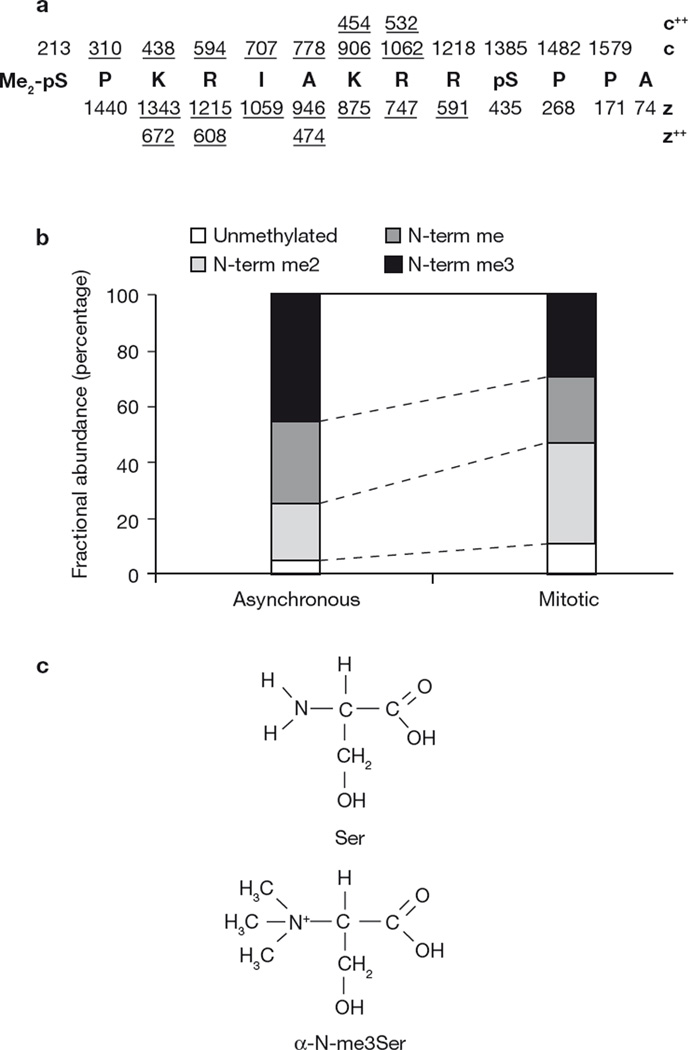
Regulator of chromatin condensation 1 (RCC1) is the only known guanine nucleotide-exchange factor for the Ran GTPase and has pivotal roles in nucleo-cytoplasmic transport, mitosis, and nuclear-envelope assembly. RCC1 associates dynamically with chromatin through binding to histones H2A and/or H2B in a Ran-regulated manner. Here, we report that, unexpectedly, the amino-terminal serine or proline residue of RCC1 is uniquely methylated on its alpha-amino group. Methylation requires removal of the initiating methionine, and the presence of proline and lysine at positions 3 and 4, respectively. Methylation-defective mutants of RCC1 bind less effectively than wild-type protein to chromatin during mitosis, which causes spindle-pole defects. We propose a bimodal attachment mechanism for RCC1 in which the tail promotes stable RCC1 association with chromatin through DNA binding in an alpha-N-methylation-dependent manner. These data provide the first known function for N-terminal protein methylation.


Figures

Figure 1
Figure 1
RCC1 N-terminal methylation. (a) Summary of ETD mass spectral data, recorded on the (M+4H)+4 ion corresponding to the 13-residue, N-terminal peptide of RCC1–Flag (residues 2–14 of the gene encoded sequence). RCC1 was isolated from mitotically arrested tsBN2 cells. Nominal m/z values for singularly charged ions of type c and z are shown above and below the peptide sequence, respectively. Those observed in the spectrum are underlined. Observed doubly charged c and z ions are also underlined. The spectra are shown in the Supplementary Information, Fig. S1a. m/z values for y2, y3 and y12 ions observed in the corresponding collision activated dissociation (CAD) mass spectrum also support the above sequence assignment (data not shown). We conclude that the peptide contains α-dimethyl- and phosphate-groups on the N-terminal-Ser 2 residue (Me2-pS2) and phosphate on Ser 11 (pS11). Experimental and calculated precursor m/z values for a peptide of this composition agree to within 2.5 p. p.m. (413.7190 and 413.7199, respectively). (b) Relative abundances of RCC1 N-terminal peptides (2–14) from asynchronous versus mitotically arrested tsBN2 cells as determined by semi-quantitative tandem mass spectrometry. Mono- and dimethylated RCC1 was also phosphorylated on Ser 2 in mitotic cells (data not shown). (c) Schematic representation of serine and me3Ser structures.
Figure 2
Figure 2
Identification of N-terminal methylation motif, α-N-terminal methyltransferase activity and detection of endogenous RCC1 N-terminal methylation. (a) Relative abundances of RCC1 N-terminal peptide (2–14) from wild type (WT), S2A, S2P, ASPK, P3Q, K4Q and K4R mutants isolated from asynchronous HeLa cells as determined by tandem mass spectrometry. RCC1ASPK–Flag and RCC1P3Q–Flag are both N-terminally acetylated. (b) N-terminal methylation of RCC1–6xHis by soluble HeLa nuclear extract. Recombinant RCC1 or buffer was incubated with extract and 3H-SAM. Incorporation of 3H-methyl group into RCC1–6xHis was detected by filter binding and scintillation counting. (c, d) Immunoblots using an anti-N-me2Ser 2 antibody (c) and anti-N-me2/3Ser 2 antibody (d). Blots are shown for recombinant RCC1 +/− methylation by nuclear extract and SAM. Antibody specificities were determined by dot blots of the indicated peptides. Uncropped images of the blots are shown in the Supplementary Information, Fig. S5. (e) Detection of α-N-methylation on endogenous RCC1. Immunoblots for HeLa cell extracts transfected with RCC1 siRNAs or a control siRNA using the indicated antibodies. Ran was used as a loading control.
Figure 3
Figure 3
Methylation of RCC1 regulates its interaction with chromosomes. (a) Mutations disrupt the localization of RCC1 in fixed mitotic MDCK cells. Cells expressing GFP or Flag fusions with either wild-type or mutant RCC1 were fixed and immunostained for tubulin and Flag (where indicated). Ratios of relative fluorescence intensity of RCC1 at chromosome versus centrosome were measured. Data were analysed by two-tailed independent t tests. Single asterisk indicates P ≤0.001, double asterisk indicates P ≤1E–08, three asterisks indicate P = 0.00011; n >30 cells per sample from three independent experiments. The error bars represent s.d. (b) Mutation disrupts the localization of RCC1 in live unfixed mitotic MDCK cells. Images of live cells expressing RCC1–GFP or RCC1K4R–GFP were collected. Sample pictures represent cells with two different protein expression levels for each construct. Ratios of relative fluorescence intensity of cytosolic versus chromatin bound RCC1 were measured. Data were analysed by two-tailed independent t tests (n ≥35 cells per sample from four independent experiments). The error bars represent s.d. (c) FRAP of RCC1–GFP in mitotic tsBN2 cells. Examples of representative cells expressing RCC1K4Q–GFP fusion proteins in metaphase are shown. The error bars represent s.d. from three independent experiments. Percentage recovery at 14 s was analysed by two-tailed independent t tests. For wild type (n = 11) versus RCC1K4Q (n = 10), P = 0.00022 and for RCC1S2A (n = 15) versus RCC1K4Q, P = 3.98E–05. (d) FLIP of RCC1–GFP in mitotic MDCK cells. Spots marked by a circle in mitotic cytosol were repeatedly photobleached. Representative FLIP images are shown. Data were analysed by 2-way ANOVA with posthoc comparisons by Tukey’s Honestly Significant Differences Test (HSD): wild type versus RCC1D182A (P <0.003, P <0.05 post hoc), wild type versus RCC1K4Q (P <0.00004, P <0.05 post hoc), wild type versus RCC1K4Q,D182A (P <0.0002, P <0.05 post hoc); n = 11 cells each with 100 time-points from three independent experiments. The scale bars in a, b and c represent 10 µm.
Figure 4
Figure 4
Methylation of RCC1 is required for correct spindle assembly and chromosome segregation. (a) Examples of cells expressing RCC1–GFP with normal spindles and chromosomes or expressing RCC1K4Q –GFP with multiple spindle pole defects are shown. (b) MDCK cells expressing RCC1–Flag, RCC1K4Q–Flag, RCC1–Flag–H2A or RCC1K4Q–FLAG–H2A were fixed and stained. (c) Methylation of RCC1 facilitates its interaction with DNA. 35S-labelled recombinant RCC1–6xHis and 3H-labelled, N-terminally methylated RCC1–6xHis were combined, then incubated with DNA agarose or biotin-labelled recombinant histone H2A on streptavidin beads, or GST–importin-α3 on glutathione–Sepharose beads, or with agarose beads alone. Beads and supernatant were separated without washing by brief centrifugation and the ratio of 35S:3H was measured by scintillation counting. Data were analysed by two-tailed independent t test, n = 3. (d) Methylation promotes binding of the RCC1 tail to mitotic chromosomes in living unfixed MDCK cells. Cells expressing the indicated constructs were stained for DNA using Hoechst dye. Chromosomal:cytosolic GFP ratios were measured and analysed by two-tailed t-test. Asterisk indicates signficant differences from wild type (n = 14) for: RCC1K4R, P = 3.54E–05 (n = 14); RCC1K4Q, P = 1.9E–05 (n = 10); RCC1ASPK, P = 1.07E–05 (n = 10). (e) Methylated recombinant RCC1 tail (1–34) binds to nuclei with higher affinity than the unmethylated protein. Recombinant RCC11–34–CFP or RCC11–34–YFP were N-terminally methylated in vitro using HeLa nuclear extract. Indicated mixture of proteins was added to permeablized MDCK cells. After incubation, cells were incubated in PBS and imaged. Data were analysed by two-tailed independent t tests, n = 3. Single asterisk indicates P = 0.048; double asterisk indicates P = 0.027. The scale bars in a, b, c and e represent 10 µm. All error bars represent s.d. Y, YFP; C, CFP.

Comment in

  • Anchoring RCC1 by the tail.
    Clarke PR. Clarke PR. Nat Cell Biol. 2007 May;9(5):485-7. doi: 10.1038/ncb0507-485. Nat Cell Biol. 2007. PMID: 17473856 No abstract available.

References

    1. Hetzer M, Gruss OJ, Mattaj IW. The Ran GTPase as a marker of chromosome position in spindle formation and nuclear envelope assembly. Nature Cell Biol. 2002;4:E177–E184. - PubMed
    1. Nemergut ME, Mizzen CA, Stukenberg T, Allis CD, Macara IG. Chromatin docking and exchange activity enhancement of RCC1 by histones H2A and H2B. Science. 2001;292:1540–1543. - PubMed
    1. Li HY, Wirtz D, Zheng Y. A mechanism of coupling RCC1 mobility to RanGTP production on the chromatin in vivo. J. Cell Biol. 2003;160:635–644. - PMC - PubMed
    1. Renault L, Kuhlmann J, Henkel A, Wittinghofer A. Structural basis for guanine nucleotide exchange on Ran by the regulator of chromosome condensation (RCC1) Cell. 2001;105:245–255. - PubMed
    1. Talcott B, Moore MS. The nuclear import of RCC1 requires a specific nuclear localization sequence receptor, karyopherin α3/Qip. J. Biol. Chem. 2000;275:10099–10104. - PubMed

Publication types

MeSH terms