. 2007 May;10(5):615-22.
doi: 10.1038/nn1876. Epub 2007 Apr 15.

Astrocytes expressing ALS-linked mutated SOD1 release factors selectively toxic to motor neurons

Affiliations

Astrocytes expressing ALS-linked mutated SOD1 release factors selectively toxic to motor neurons

Makiko Nagai et al. Nat Neurosci. 2007 May.

Abstract

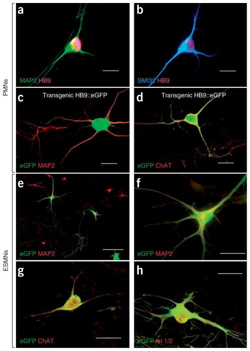
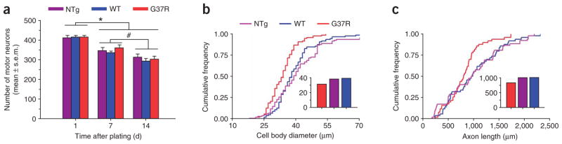
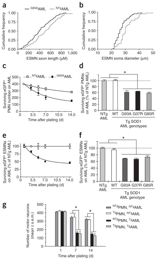
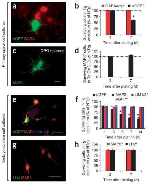
Mutations in superoxide dismutase-1 (SOD1) cause a form of the fatal paralytic disorder amyotrophic lateral sclerosis (ALS), presumably by a combination of cell-autonomous and non-cell-autonomous processes. Here, we show that expression of mutated human SOD1 in primary mouse spinal motor neurons does not provoke motor neuron degeneration. Conversely, rodent astrocytes expressing mutated SOD1 kill spinal primary and embryonic mouse stem cell-derived motor neurons. This is triggered by soluble toxic factor(s) through a Bax-dependent mechanism. However, mutant astrocytes do not cause the death of spinal GABAergic or dorsal root ganglion neurons or of embryonic stem cell-derived interneurons. In contrast to astrocytes, fibroblasts, microglia, cortical neurons and myocytes expressing mutated SOD1 do not cause overt neurotoxicity. These findings indicate that astrocytes may play a role in the specific degeneration of spinal motor neurons in ALS. Identification of the astrocyte-derived soluble factor(s) may have far-reaching implications for ALS from both a pathogenic and therapeutic standpoint.


Conflict of interest statement

COMPETING INTERESTS STATEMENT

The authors declare no competing financial interests.

Figures

Figure 1
Figure 1
Our two culture systems show well defined spinal motor neurons. (a,b) Immunostaining of primary neuronal cultures showing MAP2+HB9+ (a) and SMI32+HB9+ (b) large multipolar PMNs derived from nontransgenic mouse embryos. All were plated on NTgAML. (c,d) Double immunostaining of primary neuronal cultures showing MAP2+eGFP+ (c) and ChAT+eGFP+ (d) PMNs derived from a transgenic Hlxb9-GFP1Tmj embryo. (eh) ESMNs expressing eGFP under the HB9 promoter cultured for 7 d on spinal cord astrocyte monolayers. Confirming their motor neuron phenotype, eGFP+HB9+ ESMNs are immunopositive for MAP2 (e,f), ChAT (g) and Islet (Isl) 1/2 (h). Scale bars, 50 μm (ad,fh) and 100 μm (e).
Figure 2
Figure 2
Mutated SOD1 expressed in primary spinal-cord motor neurons provokes a mild cell-autonomous phenotype. (a) Quantification of NTg, WT and G37RPMNs at 1, 7 and 14 d after plating, showing progressive decreases (P < 0.01) in PMN numbers, but no difference among the various genotypes (P > 0.05). *Lower (P < 0.01) than day 1; different from each other (P < 0.05). Values represent means ± s.e.m. from at least three independent experiments performed at least in triplicate, analyzed by two-way ANOVA followed by a Newman-Keuls post hoc test. (b,c) At 14 d after plating, G37RPMNs show (Kolmogorov-Smirnov test; P < 0.001) smaller cell body diameters (b) and shorter axon lengths (c) than WT and NTgPMNs. Insets represent the respective medians. For these analyses, we used data from 50–100 motor neurons per genotype.
Figure 3
Figure 3
Marked toxicity of mutated SOD1–expressing astrocytes to motor neurons. (a,b) ESMNs show shorter axonal lengths (a) and smaller cell body diameters (b) when plated on AMLs expressing SOD1G93A, compared with their counterparts plated on NTgAMLs (Kolmogorov-Smirnov test; P < 0.005). (c) The decay in numbers of eGFPPMNs plated on G93AAMLs is greater (F3,37 = 6.3, P = 0.0015) than that of eGFPPMNs plated on NTgAMLs. (d) At 7 d after plating, there are consistently fewer eGFPPMNs (P < 0.01) in G93A, G37R and G85RAML cocultures than in NTgAML cocultures. However, WT and NTgAML cocultures have similar (P > 0.05) numbers of NTgPMNs. (e,f) ESMNs plated on either NTg or TgAMLs behave similarly to PMNs. (g) There are comparable numbers (P > 0.05) of NTg and G37RPMNs surviving at all time points after plating on NTgAMLs. There are fewer (*P < 0.01) NTg and G37RPMNs surviving on G93AAMLs than on NTgAMLs. The loss of G37RPMNs plated on G93AAMLs is comparable (P > 0.05) to that of NTgPMNs. Values represent means ± s.e.m. from at least three independent experiments performed at least in triplicate, analyzed by two-way ANOVA followed by a Newman-Keuls post hoc test. Tg, transgenic.
Figure 4
Figure 4
Media conditioned specifically by astrocytes expressing mutated SOD1 kill primary spinal cord and embryonic stem cell–derived motor neurons. (a) There are more eGFPPMNs surviving after 7 d of culture in fresh medium or NTgAML conditioned medium (CMd) than in G93AAML conditioned medium (*P < 0.01). (b,c) In contrast to the medium conditioned with G93AAML (*P < 0.01), media conditioned with SOD1G93A cerebral cortical neurons, skeletal myotubes or skin fibroblasts have no effect (P > 0.05) on either PMN or ESMN survival compared with controls (ESMNs cultured with medium conditioned with nontransgenic cells). Medium conditioned with spinal SOD1G93A microglia plated at a density twice that of astrocytes has only mild toxic effects on PMN (#P < 0.05) or ESMN (P > 0.05) survival. Values represent means ± s.e.m. from at least three independent experiments performed at least in triplicate, analyzed by two-way ANOVA followed by a Newman-Keuls post hoc test.
Figure 5
Figure 5
Neither mutant SOD1 AMLs nor conditioned media impair survival of neurons other than motor neurons. (a) eGFPPMNs on an AML, immunostained for eGFP and GABA at 7 d. (b) The percentage of surviving eGFPPMNs on G93AAMLs at 7 d is lower (*P < 0.004) than that at 1 d, whereas that of surviving GABAergic neurons on G93AAMLs is identical at 1 and 7 d (P > 0.5). (c,d) The percentages of surviving MAP2+ DRG neurons cultured with G93AAML conditioned medium for 2 d or for 7 d are also identical (P > 0.05). (e) Within the same culture, among the embryonic stem cell–derived MAP2+ neurons, some are eGFP+HB9+ (ESMNs) and others are eGFPHB9; among the latter, some are Lim1/2+. (f) Five days after embryonic stem cell–derived neurons were plated, the percentage of surviving ESMNs on G93AAMLs is lower (*P < 0.01) than that on NTgAMLs. In contrast, the percentages of neither MAP2+eGFPHB9(MAP2+GFP) (F3,24 = 0.4, P = 0.8) nor MAP2+eGFPHB9Lim1/2+ (Lim1/2+) neurons (F3,16 = 0.1, P = 0.9) differs between the two AML cocultures. (g) Embryonic stem cell–derived neurons differentiated into posterior interneurons expressing the LH2 marker. (h) The percentages of surviving LH2+ or MAP2+ neurons are not different on NTg or G93AAMLs at 2 or 7 d (P > 0.05). Values represent means ± s.e.m. from at least three independent experiments performed at least in triplicate, analyzed by two-way ANOVA followed by a Newman-Keuls post hoc test. Scale bars, 100 μm (a), 20 μm (c) and 50 μm (e,g).
Figure 6
Figure 6
ESMNs die in response to mutant AMLs through a Bax-dependent mechanism. (ae) Death of ESMNs assessed by immunostaining for fractin and by EthD uptake. At 7 d after plating, the Bax inhibitor V5 decreases (P < 0.01) the percentages of ESMNs labeled with EthD (a) and embryonic stem cell–derived MAP2+fractin+ neurons (d) and increases (*P < 0.01) the percentages of surviving ESMNs (e). Values are expressed as percentage of NTgAML-coculture values and represent means ± s.e.m. from at least three independent experiments performed at least in triplicate, analyzed by two-way ANOVA followed by a Newman-Keuls post hoc test. *Higher (P < 0.01) than NTgAML cocultures; #lower (P < 0.001) than TgAML cocultures with vehicle (veh). (b,c) All ESMNs immunopositive for fractin show DNA condensations, as evidenced by Hoechst 33342 (b), and all ESMNs with Hoechst 33342–labeled chromatin clumps show DNA fragmentation, as evidenced by TUNEL (c). Scale bar, 20 μm.

Comment in

References

    1. Rosen DR, et al. Mutations in Cu/Zn superoxide dismutase gene are associated with familial amyotrophic lateral sclerosis. Nature. 1993;362:59–62. - PubMed
    1. Deng HX, et al. Amyotrophic lateral sclerosis and structural defects in Cu,Zn superoxide dismutase. Science. 1993;261:1047–1051. - PubMed
    1. Gurney ME, et al. Motor neuron degeneration in mice that express a human Cu, Zn superoxide dismutase mutation. Science. 1994;264:1772–1775. - PubMed
    1. Wong PC, et al. An adverse property of a familial ALS-linked SOD1 mutation causes motor neuron disease characterized by vacuolar degeneration of mitochondria. Neuron. 1995;14:1105–1116. - PubMed
    1. Bruijn LI, et al. ALS-linked SOD1 mutant G85R mediated damage to astrocytes and promotes rapidly progressive disease with SOD1-containing inclusions. Neuron. 1997;18:327–338. - PubMed

Publication types

MeSH terms