Practice Guideline
. 2007 May;114(3):111-6.
doi: 10.1007/s10633-007-9053-1. Epub 2007 Apr 14.

ISCEV standard for clinical pattern electroretinography--2007 update

Affiliations
Practice Guideline

ISCEV standard for clinical pattern electroretinography--2007 update

Graham E Holder et al. Doc Ophthalmol. 2007 May.

Abstract

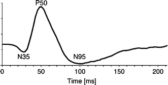
The pattern electroretinogram (PERG) is a retinal response evoked by viewing a temporally alternating pattern, usually a black and white checkerboard or grating. The PERG is important in clinical and research applications because it provides information both about retinal ganglion cell function and, because the stimulus is customarily viewed with central fixation, the function of the macula. The PERG can therefore facilitate interpretation of an abnormal pattern VEP by revealing the retinal responses to a similar stimulus to that used for the VEP. However, practitioners may have difficulty choosing between the different techniques for recording the PERG that have been described in the literature. The International Society for Clinical Electrophysiology of Vision published a standard for clinical PERG recording in 2000 to assist practitioners in obtaining good quality reliable responses and to facilitate inter-laboratory communication and comparison. This document is the scheduled revision of that standard.


Figures

Fig. 1
Fig. 1
A normal PERG. The amplitude of P50 in a normal subject is usually between 2.0 and 4.0 μV

References

    1. {'text': '', 'ref_index': 1, 'ids': [{'type': 'DOI', 'value': '10.1007/BF01214647', 'is_inner': False, 'url': 'https://doi.org/10.1007/bf01214647'}, {'type': 'PubMed', 'value': '8899299', 'is_inner': True, 'url': 'https://pubmed.ncbi.nlm.nih.gov/8899299/'}]}
    2. Marmor MF, Holder GE, Porciatti V, Trick GL, Zrenner E (1996) Guidelines for basic pattern electroretinography. Recommendations by the International Society for Clinical Electrophysiology of Vision. Doc Ophthalmol 91:291–298 - PubMed
    1. {'text': '', 'ref_index': 1, 'ids': [{'type': 'DOI', 'value': '10.1023/A:1002732114721', 'is_inner': False, 'url': 'https://doi.org/10.1023/a:1002732114721'}, {'type': 'PubMed', 'value': '11128964', 'is_inner': True, 'url': 'https://pubmed.ncbi.nlm.nih.gov/11128964/'}]}
    2. Bach M, Hawlina M, Holder GE, Marmor MF, Meigen T, Vaegan, Miyake Y (2000) Standard for Pattern Electroretinography. Doc Ophthalmol 101:11–18 - PubMed
    1. {'text': '', 'ref_index': 1, 'ids': [{'type': 'DOI', 'value': '10.1023/A:1001724411607', 'is_inner': False, 'url': 'https://doi.org/10.1023/a:1001724411607'}, {'type': 'PubMed', 'value': '10189178', 'is_inner': True, 'url': 'https://pubmed.ncbi.nlm.nih.gov/10189178/'}]}
    2. Brigell M, Bach M, Barber C, Kawasaki K, Kooijman A (1998) Guidelines for calibration of stimulus and recording parameters used in clinical electrophysiology of vision. Doc Ophthalmol 95:1–14 - PubMed
    1. {'text': '', 'ref_index': 1, 'ids': [{'type': 'DOI', 'value': '10.1023/B:DOOP.0000036793.44912.45', 'is_inner': False, 'url': 'https://doi.org/10.1023/b:doop.0000036793.44912.45'}, {'type': 'PubMed', 'value': '15455793', 'is_inner': True, 'url': 'https://pubmed.ncbi.nlm.nih.gov/15455793/'}]}
    2. Marmor MF, Holder GE, Seeliger MW, Yamamoto S (2004) Standard for clinical electroretinography (2004 update). Doc Ophthalmol 108:107–114 - PubMed

Publication types

MeSH terms