

Biodegradable poly(2-dimethylamino ethylamino)phosphazene for in vivo gene delivery to tumor cells. Effect of polymer molecular weight
- PMID: 17435970
- PMCID: PMC1915646
- DOI: 10.1007/s11095-007-9299-z
Biodegradable poly(2-dimethylamino ethylamino)phosphazene for in vivo gene delivery to tumor cells. Effect of polymer molecular weight
Abstract
Purpose: Previously, we have shown that complexes of plasmid DNA with the biodegradable polymer poly(2-dimethylamino ethylamino)phosphazene (p(DMAEA)-ppz) mediated tumor selective gene expression after intravenous administration in mice. In this study, we investigated the effect of p(DMAEA)-ppz molecular weight on both in vitro and in vivo tumor transfection, as well as on complex induced toxicity.
Materials and methods: p(DMAEA)-ppz with a broad molar mass distribution was fractionated by preparative size exclusion chromatography. Polyplexes consisting of plasmid DNA and the collected polymer fractions were tested for biophysical properties, (cyto)toxicity and transfection activity.
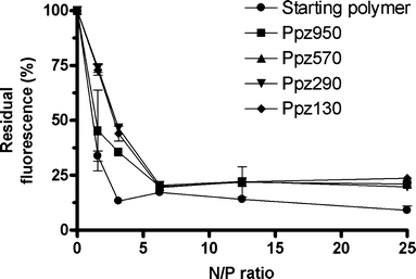
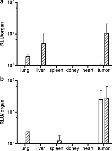
Results: Four p(DMAEA)-ppz fractions were collected with weight average molecular weights ranging from 130 to 950 kDa, and with narrow molecular mass distributions (Mw/Mn from 1.1 to 1.3). At polymer-to-DNA (N/P) ratios above 6, polyplexes based on these polymers were all positively charged (zeta potential 25-29 mV), and had a size of 80-90 nm. The in vitro cytotoxicity of the polyplexes positively correlated with polymer molecular weight. The in vitro transfection activity of the different polyplexes depended on their N/P ratio, and was affected by the degree of cytotoxicity, as well as the colloidal stability of the different polyplexes. Intravenous administration of polyplexes based on the high molecular weight polymers led to apparent toxicity, as a result of polyplex-induced erythrocyte aggregation. On the other hand, administration of polyplexes based on low molecular weight p(DMAEA)-ppz's (Mw 130 kDa) did not show signs of toxicity and resulted in tumor selective gene expression.
Conclusion: Polymer molecular weight fractionation enabled us to optimize the transfection efficiency/toxicity ratio of p(DMAEA)-ppz polyplexes for in vitro and in vivo tumor transfection.
Figures


Similar articles
-
In vivo tumor transfection mediated by polyplexes based on biodegradable poly(DMAEA)-phosphazene.J Control Release. 2005 Dec 5;109(1-3):275-87. doi: 10.1016/j.jconrel.2005.05.030. Epub 2005 Jul 21. J Control Release. 2005. PMID: 16039747
-
Degradable PEG-folate coated poly(DMAEA-co-BA)phosphazene-based polyplexes exhibit receptor-specific gene expression.Eur J Pharm Sci. 2008 Mar 3;33(3):241-51. doi: 10.1016/j.ejps.2007.12.003. Epub 2007 Dec 8. Eur J Pharm Sci. 2008. PMID: 18207707
-
Polyplex formation between four-arm poly(ethylene oxide)-b-poly(2-(diethylamino)ethyl methacrylate) and plasmid DNA in gene delivery.J Biomed Mater Res A. 2009 Dec;91(3):708-18. doi: 10.1002/jbm.a.32255. J Biomed Mater Res A. 2009. PMID: 19048636
-
Targeted polymers for gene delivery.Expert Opin Drug Deliv. 2005 Jan;2(1):145-57. doi: 10.1517/17425247.2.1.145. Expert Opin Drug Deliv. 2005. PMID: 16296741 Review.
-
Gene delivery by lipoplexes and polyplexes.Eur J Pharm Sci. 2010 Jun 14;40(3):159-70. doi: 10.1016/j.ejps.2010.03.019. Epub 2010 Mar 30. Eur J Pharm Sci. 2010. PMID: 20359532 Review.
Cited by
-
Reducible HPMA-co-oligolysine copolymers for nucleic acid delivery.Int J Pharm. 2012 May 1;427(1):113-22. doi: 10.1016/j.ijpharm.2011.08.015. Epub 2011 Aug 27. Int J Pharm. 2012. PMID: 21893178 Free PMC article.
-
Engineering biodegradable and multifunctional peptide-based polymers for gene delivery.J Biol Eng. 2013 Oct 24;7(1):25. doi: 10.1186/1754-1611-7-25. J Biol Eng. 2013. PMID: 24156736 Free PMC article.
-
Increased RNAi Efficacy in Spodoptera exigua via the Formulation of dsRNA With Guanylated Polymers.Front Physiol. 2018 Apr 4;9:316. doi: 10.3389/fphys.2018.00316. eCollection 2018. Front Physiol. 2018. PMID: 29670535 Free PMC article.
-
Cathepsin B-sensitive polymers for compartment-specific degradation and nucleic acid release.J Control Release. 2012 Feb 10;157(3):445-54. doi: 10.1016/j.jconrel.2011.10.016. Epub 2011 Oct 20. J Control Release. 2012. PMID: 22036879 Free PMC article.
-
Polyphosphazene-Based Nanotherapeutics.J Funct Biomater. 2025 Aug 2;16(8):285. doi: 10.3390/jfb16080285. J Funct Biomater. 2025. PMID: 40863305 Free PMC article. Review.
References
-
- {'text': '', 'ref_index': 1, 'ids': [{'type': 'DOI', 'value': '10.1097/00001813-200104000-00001', 'is_inner': False, 'url': 'https://doi.org/10.1097/00001813-200104000-00001'}, {'type': 'PubMed', 'value': '11335785', 'is_inner': True, 'url': 'https://pubmed.ncbi.nlm.nih.gov/11335785/'}]}
- A. G. Schatzlein. Non-viral vectors in cancer gene therapy: principles and progress. Anticancer Drugs12:275–304 (2001). - PubMed
-
- {'text': '', 'ref_index': 1, 'ids': [{'type': 'DOI', 'value': '10.1016/S1359-6446(02)02243-2', 'is_inner': False, 'url': 'https://doi.org/10.1016/s1359-6446(02)02243-2'}, {'type': 'PubMed', 'value': '11965397', 'is_inner': True, 'url': 'https://pubmed.ncbi.nlm.nih.gov/11965397/'}]}
- M. Ogris, and E. Wagner. Targeting tumors with non-viral gene delivery systems. Drug Discov. Today7:479–485 (2002). - PubMed
-
- {'text': '', 'ref_index': 1, 'ids': [{'type': 'DOI', 'value': '10.1016/S0169-409X(02)00228-4', 'is_inner': False, 'url': 'https://doi.org/10.1016/s0169-409x(02)00228-4'}, {'type': 'PubMed', 'value': '12628320', 'is_inner': True, 'url': 'https://pubmed.ncbi.nlm.nih.gov/12628320/'}]}
- J. Panyam, and V. Labhasetwar. Biodegradable nanoparticles for drug and gene delivery to cells and tissue. Adv. Drug Deliv. Rev. 55:329–347 (2003). - PubMed
-
- {'text': '', 'ref_index': 1, 'ids': [{'type': 'DOI', 'value': '10.1016/S1367-5931(00)00227-1', 'is_inner': False, 'url': 'https://doi.org/10.1016/s1367-5931(00)00227-1'}, {'type': 'PubMed', 'value': '11470609', 'is_inner': True, 'url': 'https://pubmed.ncbi.nlm.nih.gov/11470609/'}]}
- O. Pillai, and R. Panchagnula. Polymers in drug delivery. Curr. Opin. Chem. Biol. 5:447–451 (2001). - PubMed
-
- {'text': '', 'ref_index': 1, 'ids': [{'type': 'DOI', 'value': '10.1016/j.jconrel.2005.05.030', 'is_inner': False, 'url': 'https://doi.org/10.1016/j.jconrel.2005.05.030'}, {'type': 'PubMed', 'value': '16039747', 'is_inner': True, 'url': 'https://pubmed.ncbi.nlm.nih.gov/16039747/'}]}
- H. K. De Wolf, J. Luten, C. J. Snel, C. Oussoren, W. E. Hennink, and G. Storm. In vivo tumor transfection mediated by polyplexes based on biodegradable poly(DMAEA)-phosphazene. J. Control. Release109:275–287 (2005). - PubMed
MeSH terms
Substances
LinkOut - more resources
Full Text Sources
Other Literature Sources
Miscellaneous
