

Management of hepatic angiomyolipoma
- PMID: 17436129
- PMCID: PMC1852378
- DOI: 10.1007/s11605-006-0037-3
Management of hepatic angiomyolipoma
Abstract
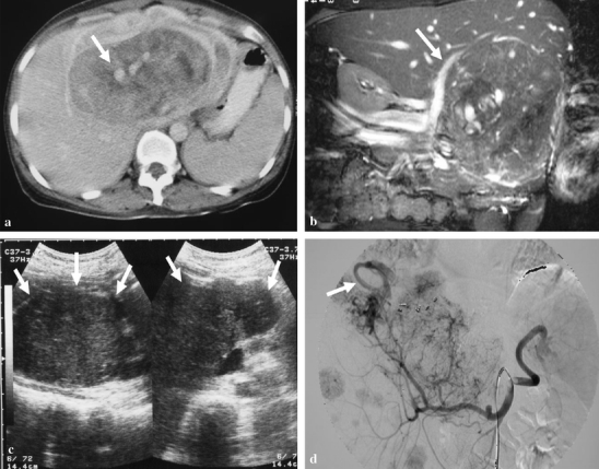
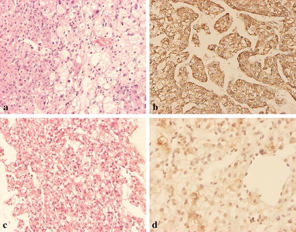
Preoperative diagnosis of hepatic angiomyolipoma is difficult, and the treatment for it remains controversial. The aim of this study is to review our experience in the treatment of hepatic angiomyolipoma and to propose a treatment strategy for this disease. We retrospectively collected the clinical, imaging, and pathological features of patients with hepatic angiomyolipoma. Immunohistochemical studies with antibodies for HMB-45, actin, S-100, cytokeratin, vimentin, and c-kit were performed. Treatment experience and long-term follow-up results are summarized. During a period of 9 years, 10 patients with hepatic angiomyolipoma were treated at our hospital. There was marked female predominance (nine patients). Nine patients received surgical resection without complications. One patient received nonoperative management with biopsy and follow-up. One patient died 11 months after surgery because of recurrent disease. We propose all symptomatic patients should receive surgical resection for hepatic angiomyolipoma. Conservative management with close follow-up is suggested in patients with asymptomatic tumors and meet the following criteria: (1) tumor size smaller than 5 cm, (2) angiomyolipoma proved through fine needle aspiration biopsy, (3) patients with good compliance, and (4) not a hepatitis virus carrier.
Figures
References
-
- None
- Ishak KG. Mesenchymal tumors of the liver. In: Okuda K, Peters RL, eds. Hepatocellular carcinoma. New York: John Wiley and Sons:, 1976 247–307.
-
- {'text': '', 'ref_index': 1, 'ids': [{'type': 'DOI', 'value': '10.1016/j.jpedsurg.2004.08.036', 'is_inner': False, 'url': 'https://doi.org/10.1016/j.jpedsurg.2004.08.036'}, {'type': 'PubMed', 'value': '15616929', 'is_inner': True, 'url': 'https://pubmed.ncbi.nlm.nih.gov/15616929/'}]}
- Kitano Y, Honna T, Nihei K, et al. Renal angiomyolipoma in Japanese tuberous sclerosis patients. J Pediatr Surg 2004;39:1784–6. - PubMed
-
- {'text': '', 'ref_index': 1, 'ids': [{'type': 'PubMed', 'value': '11800647', 'is_inner': True, 'url': 'https://pubmed.ncbi.nlm.nih.gov/11800647/'}]}
- Makhlouf HR, Ishak KG, Shekar R, et al. Melanoma markers in angiomyolipoma of the liver and kidney: a comparative study. Arch Pathol Lab Med 2002;126:49–55. - PubMed
-
- {'text': '', 'ref_index': 1, 'ids': [{'type': 'PubMed', 'value': '8147221', 'is_inner': True, 'url': 'https://pubmed.ncbi.nlm.nih.gov/8147221/'}]}
- Ma TK, Tse MK, Tsui WM, et al. Fine needle aspiration diagnosis of angiomyolipoma of the liver using a cell block with immunohistochemical study. A case report. Acta Cytol 1994;38:257–60. - PubMed
-
- {'text': '', 'ref_index': 1, 'ids': [{'type': 'DOI', 'value': '10.1002/(SICI)1097-0142(19990225)87:1<25::AID-CNCR5>3.0.CO;2-I', 'is_inner': False, 'url': 'https://doi.org/10.1002/(sici)1097-0142(19990225)87:1<25::aid-cncr5>3.0.co;2-i'}, {'type': 'PubMed', 'value': '10096356', 'is_inner': True, 'url': 'https://pubmed.ncbi.nlm.nih.gov/10096356/'}]}
- Cha I, Cartwright D, Guis M, et al. Angiomyolipoma of the liver in fine-needle aspiration biopsies: its distinction from hepatocellular carcinoma. Cancer 1999;87:25–30. - PubMed
MeSH terms
LinkOut - more resources
Full Text Sources
Medical
