

Experimental infection with Haemophilus ducreyi in persons who are infected with HIV does not cause local or augment systemic viral replication
- PMID: 17436224
- PMCID: PMC2571042
- DOI: 10.1086/513877
Experimental infection with Haemophilus ducreyi in persons who are infected with HIV does not cause local or augment systemic viral replication
Abstract
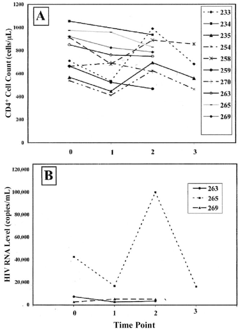
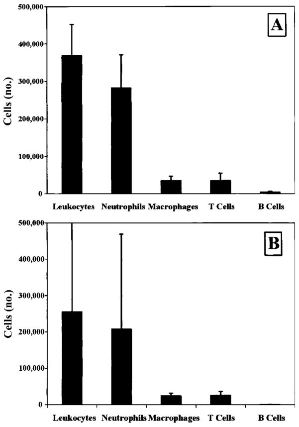
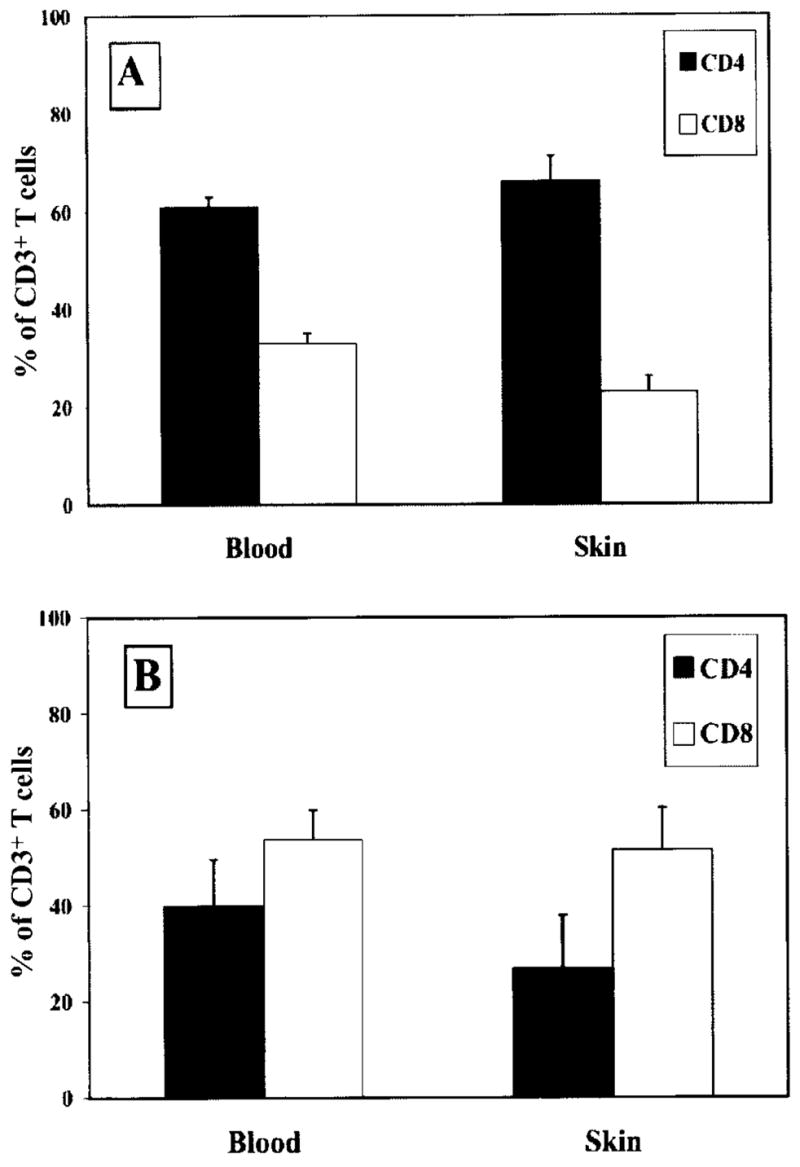
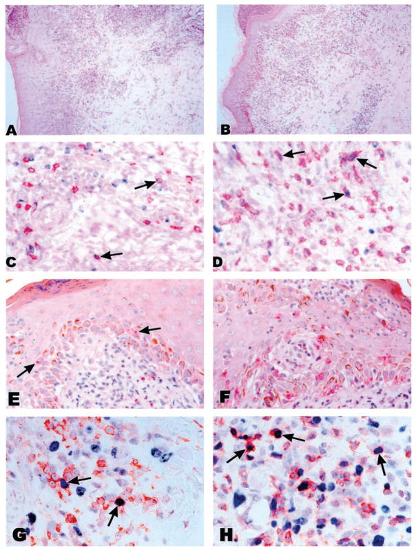
We infected 11 HIV-seropositive volunteers whose CD4(+) cell counts were >350 cells/ microL (7 of whom were receiving antiretrovirals) with Haemophilus ducreyi. The papule and pustule formation rates were similar to those observed in HIV-seronegative historical control subjects. No subject experienced a sustained change in CD4(+) cell count or HIV RNA level. The cellular infiltrate in biopsy samples obtained from the HIV-seropositive and HIV-seronegative subjects did not differ with respect to the percentage of leukocytes, neutrophils, macrophages, or T cells. The CD4(+):CD8(+) cell ratio in biopsy samples from the HIV-seropositive subjects was 1:3, the inverse of the ratio seen in the HIV-seronegative subjects (P<.0001). Although CD4(+) cells proliferated in lesions, in situ hybridization and reverse-transcription polymerase chain reaction for HIV RNA was negative. We conclude that experimental infection in HIV-seropositive persons is clinically similar to infection in HIV-seronegative persons and does not cause local or augment systemic viral replication. Thus, prompt treatment of chancroid may abrogate increases in viral replication associated with natural disease.
Conflict of interest statement
Potential conflicts of interest: none reported.
Figures
Similar articles
-
Clinical and in situ cellular responses to Haemophilus ducreyi in the presence or absence of HIV infection.Int J STD AIDS. 1998 Sep;9(9):531-6. doi: 10.1258/0956462981922773. Int J STD AIDS. 1998. PMID: 9764937
-
Enzyme immunoassays (EIAs) for the detection of anti-Haemophilus ducreyi serum IgA, IgG, and IgM antibodies.Sex Transm Dis. 1994 Jan-Feb;21(1):36-42. doi: 10.1097/00007435-199401000-00008. Sex Transm Dis. 1994. PMID: 8140487
-
Failure of treatment for chancroid in Rwanda is not related to human immunodeficiency virus infection: in vitro resistance of Haemophilus ducreyi to trimethoprim-sulfamethoxazole.Clin Infect Dis. 1995 Apr;20(4):924-30. doi: 10.1093/clinids/20.4.924. Clin Infect Dis. 1995. PMID: 7795096 Clinical Trial.
-
Low blood CD8+ T-lymphocytes and high circulating monocytes are predictors of HIV-1-associated progressive encephalopathy in children.Pediatrics. 2003 Feb;111(2):E168-75. doi: 10.1542/peds.111.2.e168. Pediatrics. 2003. PMID: 12563091
-
In vitro stimulation of peripheral blood mononuclear cells (PBMC) from HIV- and HIV+ chancroid patients by Haemophilus ducreyi antigens.Clin Exp Immunol. 1995 Nov;102(2):243-50. doi: 10.1111/j.1365-2249.1995.tb03772.x. Clin Exp Immunol. 1995. PMID: 7586673 Free PMC article.
Cited by
-
Experimental infection of human volunteers with Haemophilus ducreyi: fifteen years of clinical data and experience.J Infect Dis. 2009 Jun 1;199(11):1671-9. doi: 10.1086/598966. J Infect Dis. 2009. PMID: 19432549 Free PMC article.
-
Dysregulated immune profiles for skin and dendritic cells are associated with increased host susceptibility to Haemophilus ducreyi infection in human volunteers.Infect Immun. 2007 Dec;75(12):5686-97. doi: 10.1128/IAI.00777-07. Epub 2007 Sep 24. Infect Immun. 2007. PMID: 17893130 Free PMC article.
-
Role played by CD4+FOXP3+ regulatory T Cells in suppression of host responses to Haemophilus ducreyi during experimental infection of human volunteers.J Infect Dis. 2010 Jun 15;201(12):1839-48. doi: 10.1086/652781. J Infect Dis. 2010. PMID: 20443736 Free PMC article.
-
Host Polymorphisms in TLR9 and IL10 Are Associated With the Outcomes of Experimental Haemophilus ducreyi Infection in Human Volunteers.J Infect Dis. 2016 Aug 1;214(3):489-95. doi: 10.1093/infdis/jiw164. Epub 2016 Apr 27. J Infect Dis. 2016. PMID: 27122592 Free PMC article.
-
Haemophilus ducreyi partially activates human myeloid dendritic cells.Infect Immun. 2007 Dec;75(12):5678-85. doi: 10.1128/IAI.00702-07. Epub 2007 Oct 8. Infect Immun. 2007. PMID: 17923525 Free PMC article.
References
-
- Hayes RJ, Schulz KF, Plummer FA. The cofactor effect of genital ulcers on the per-exposure risk of HIV transmission in sub-Saharan Africa. J Trop Med Hyg. 1995;98:1–8. - PubMed
-
- Cohen MS. Thomas Parran Award Lecture: transmission and prevention of transmission of HIV-1. Sex Transm Dis. 2006;33:338–41. - PubMed
-
- Orroth KK, White RG, Korenromp EL, et al. Empirical observations underestimate the proportion of human immunodeficiency virus infections attributable to sexually transmitted diseases in the Mwanza and Rakai sexually transmitted disease treatment trials: simulation results. Sex Transm Dis. 2006;33:536–44. - PubMed
-
- Robinson NJ, Mulder DW, Auvert B, Hayes RJ. Proportion of HIV infections attributable to other sexually transmitted diseases in a rural Ugandan population: simulation model estimates. Int J Epidemiol. 1997;26:180–9. - PubMed
Publication types
MeSH terms
Substances
Grants and funding
LinkOut - more resources
Full Text Sources
Medical
Research Materials
