

Therapeutic effect of a new immunosuppressive agent, everolimus, on interleukin-10 gene-deficient mice with colitis
- PMID: 17437423
- PMCID: PMC1868878
- DOI: 10.1111/j.1365-2249.2007.03345.x
Therapeutic effect of a new immunosuppressive agent, everolimus, on interleukin-10 gene-deficient mice with colitis
Abstract
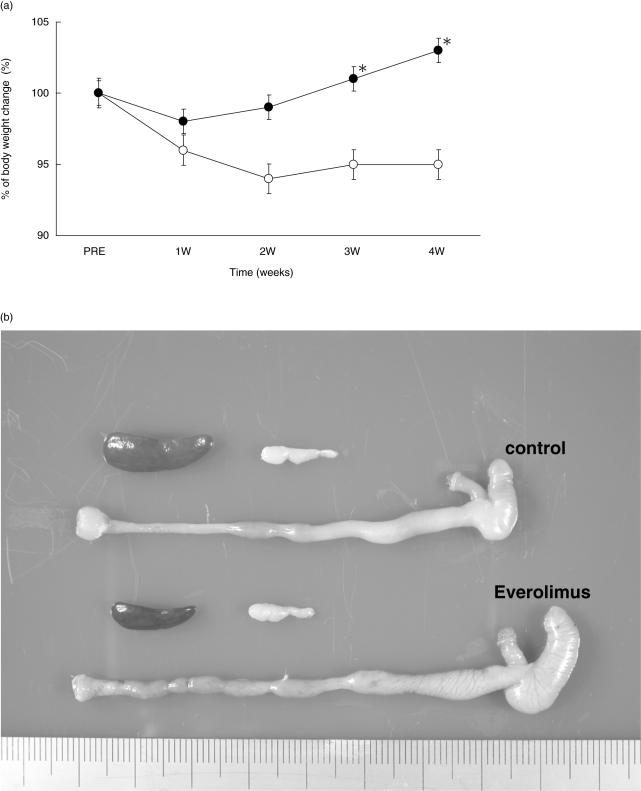
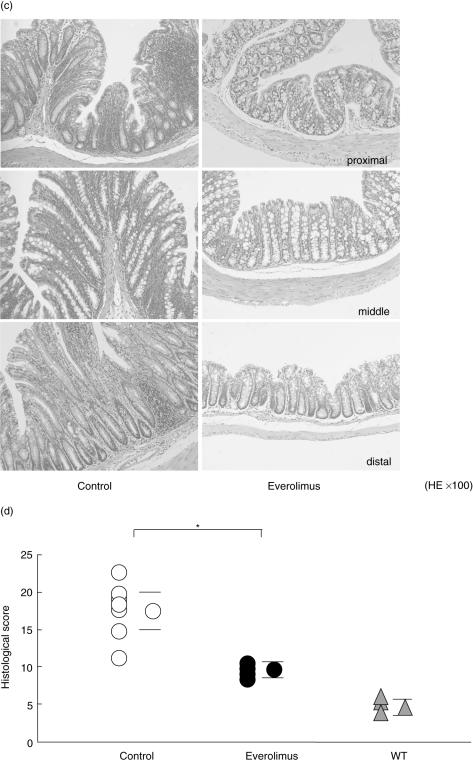
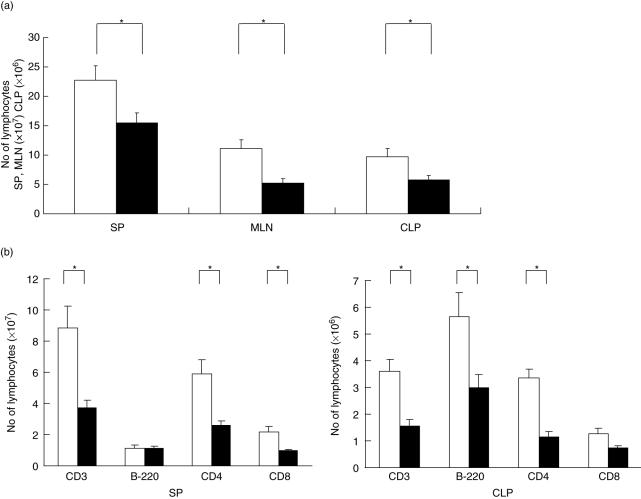
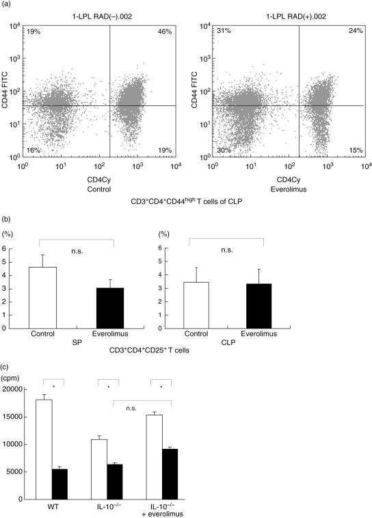
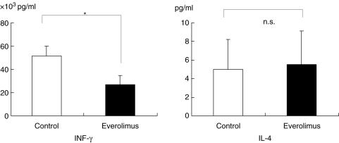
A limited number of therapeutic strategies are currently available for patients with inflammatory bowel disease (IBD). In particular, the maintenance therapy after remission in Crohn's disease (CD) is not satisfactory and new approaches are needed. Interleukin-10 gene-deficient (IL-10-/-) mice, a well-characterized experimental model of CD, develop severe chronic colitis due to an aberrant Th1 immune response. Everolimus, an inhibitor of the mammalian target of rapamycin (mTOR), a new immunosuppressive reagent, has been used successfully in animal models for heart, liver, lung and kidney transplantation. In the present study, we examined the efficacy of everolimus in the treatment of chronic colitis in an IL-10-/- mouse model. Everolimus was administered orally for a period of 4 weeks to IL-10-/- mice with clinical signs of colitis. The gross and histological appearances of the colon and the numbers, phenotype and cytokine production of lymphocytes were compared with these characteristics in a control group. The 4-week administration of everolimus resulted in a significant decrease in the severity of colitis, together with a significant reduction in the number of CD4+ T cells in the colonic lamina propria as well as IFN-gamma production in colonic lymphocytes. Everolimus treatment of established colitis in IL-10-/- mice ameliorated the colitis, probably as a result of decreasing the number of CD4+ T cells in the colonic mucosa and an associated reduction in IFN-gamma production.
Figures
Similar articles
-
Therapeutic effects of triptolide on interleukin-10 gene-deficient mice with colitis.Int Immunopharmacol. 2008 Dec 20;8(13-14):1808-12. doi: 10.1016/j.intimp.2008.08.019. Epub 2008 Sep 17. Int Immunopharmacol. 2008. PMID: 18804190
-
Therapeutic effects of a new lymphocyte homing reagent FTY720 in interleukin-10 gene-deficient mice with colitis.Inflamm Bowel Dis. 2004 May;10(3):182-92. doi: 10.1097/00054725-200405000-00002. Inflamm Bowel Dis. 2004. PMID: 15290910
-
Disparate CD4+ lamina propria (LP) lymphokine secretion profiles in inflammatory bowel disease. Crohn's disease LP cells manifest increased secretion of IFN-gamma, whereas ulcerative colitis LP cells manifest increased secretion of IL-5.J Immunol. 1996 Aug 1;157(3):1261-70. J Immunol. 1996. PMID: 8757634
-
Biodegradable microspheres targeting mucosal immune-regulating cells: new approach for treatment of inflammatory bowel disease.J Gastroenterol. 2003 Mar;38 Suppl 15:59-62. J Gastroenterol. 2003. PMID: 12698874 Review.
-
Lessons from genetically engineered animal models. XII. IL-10-deficient (IL-10(-/-) mice and intestinal inflammation.Am J Physiol Gastrointest Liver Physiol. 2000 Jun;278(6):G829-33. doi: 10.1152/ajpgi.2000.278.6.G829. Am J Physiol Gastrointest Liver Physiol. 2000. PMID: 10859210 Review.
Cited by
-
mTOR Inhibition Attenuates Dextran Sulfate Sodium-Induced Colitis by Suppressing T Cell Proliferation and Balancing TH1/TH17/Treg Profile.PLoS One. 2016 Apr 29;11(4):e0154564. doi: 10.1371/journal.pone.0154564. eCollection 2016. PLoS One. 2016. PMID: 27128484 Free PMC article.
-
Comparison of anti-inflammatory mechanisms of mango (Mangifera Indica L.) and pomegranate (Punica Granatum L.) in a preclinical model of colitis.Mol Nutr Food Res. 2016 Sep;60(9):1912-23. doi: 10.1002/mnfr.201501008. Epub 2016 May 23. Mol Nutr Food Res. 2016. PMID: 27028006 Free PMC article.
-
Targeting Immune Cell Metabolism in the Treatment of Inflammatory Bowel Disease.Inflamm Bowel Dis. 2021 Oct 18;27(10):1684-1693. doi: 10.1093/ibd/izab024. Inflamm Bowel Dis. 2021. PMID: 33693743 Free PMC article. Review.
-
Modulating T Cell Responses via Autophagy: The Intrinsic Influence Controlling the Function of Both Antigen-Presenting Cells and T Cells.Front Immunol. 2018 Dec 14;9:2914. doi: 10.3389/fimmu.2018.02914. eCollection 2018. Front Immunol. 2018. PMID: 30619278 Free PMC article. Review.
-
New insights into the interplay between autophagy, gut microbiota and inflammatory responses in IBD.Autophagy. 2020 Jan;16(1):38-51. doi: 10.1080/15548627.2019.1635384. Epub 2019 Jul 9. Autophagy. 2020. PMID: 31286804 Free PMC article. Review.
References
-
- Shanahan F. Crohn's disease. Lancet. 2002;359(9300):62–9. - PubMed
-
- Scribano ML, Prantera C. Review article: medical treatment of active Crohn's disease. Aliment Pharmacol Ther. 2002;16(Suppl. 4):35–9. - PubMed
-
- Rizzello F, Gionchetti P, Venturi A, Morselli C, Campieri M. Review article: the management of refractory Crohn's disease. Aliment Pharmacol Ther. 2002;16(Suppl. 4):40–7. - PubMed
-
- Forbes A. Review article: Crohn's disease – the role of nutritional therapy. Aliment Pharmacol Ther. 2002;16(Suppl. 4):48–52. - PubMed
-
- Sandborn WJ. A review of immune modifier therapy for inflammatory bowel disease: azathioprine, 6-mercaptopurine, cyclosporine, and methotrexate. Am J Gastroenterol. 1996;91(3):423–33. - PubMed
MeSH terms
Substances
LinkOut - more resources
Full Text Sources
Medical
Research Materials
Miscellaneous
