. 2007 Apr;15(4):395-404.
doi: 10.1016/j.str.2007.03.001.

A left-handed RNA double helix bound by the Z alpha domain of the RNA-editing enzyme ADAR1

Affiliations

A left-handed RNA double helix bound by the Z alpha domain of the RNA-editing enzyme ADAR1

Diana Placido et al. Structure. 2007 Apr.

Abstract

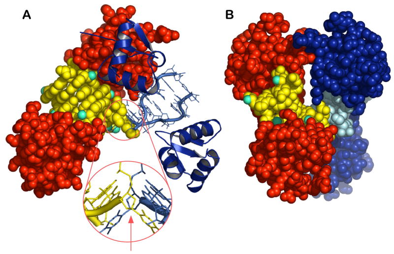
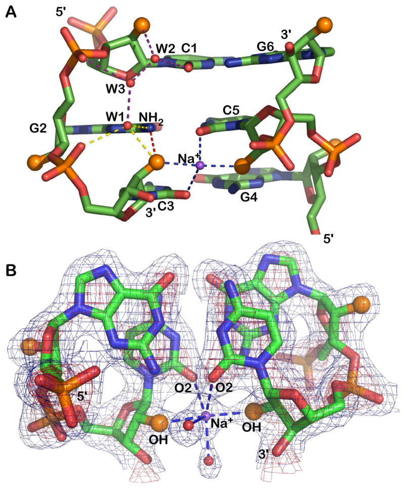
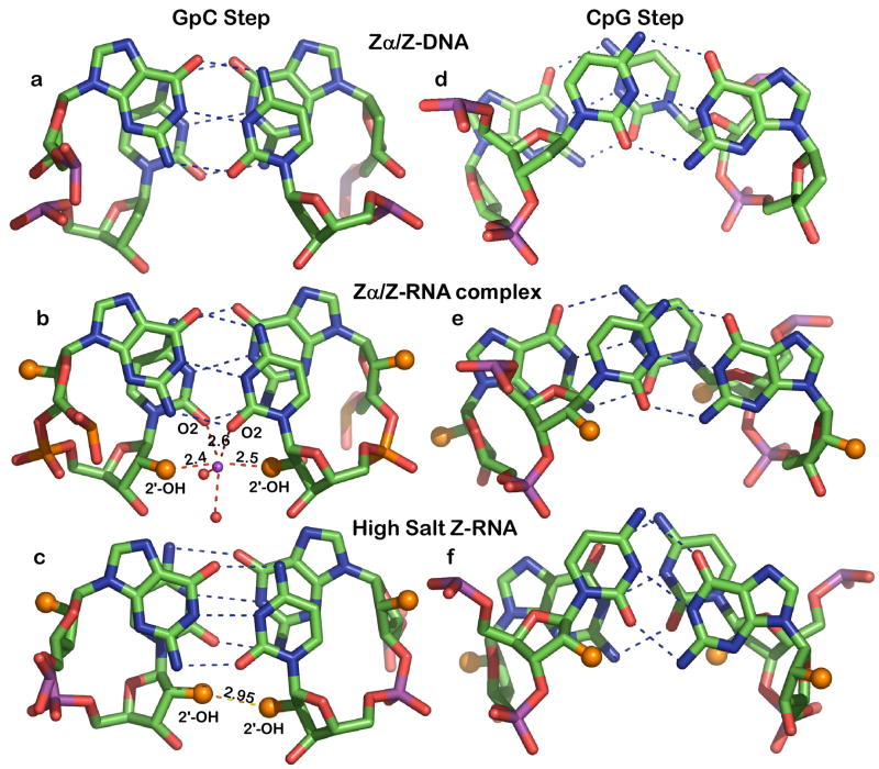
The A form RNA double helix can be transformed to a left-handed helix, called Z-RNA. Currently, little is known about the detailed structural features of Z-RNA or its involvement in cellular processes. The discovery that certain interferon-response proteins have domains that can stabilize Z-RNA as well as Z-DNA opens the way for the study of Z-RNA. Here, we present the 2.25 A crystal structure of the Zalpha domain of the RNA-editing enzyme ADAR1 (double-stranded RNA adenosine deaminase) complexed to a dUr(CG)(3) duplex RNA. The Z-RNA helix is associated with a unique solvent pattern that distinguishes it from the otherwise similar conformation of Z-DNA. Based on the structure, we propose a model suggesting how differences in solvation lead to two types of Z-RNA structures. The interaction of Zalpha with Z-RNA demonstrates how the interferon-induced isoform of ADAR1 could be targeted toward selected dsRNAs containing purine-pyrimidine repeats, possibly of viral origin.


Figures

Figure 1
Figure 1. Overall structure of the Zα/Z-RNA complex
A) The asymmetric unit has two non-interacting Zα monomers (red space-filling model) bound to a dUr(CG)3 Z-RNA duplex. A second symmetry related complex is displayed as structural motifs. The overhanging deoxy-uridine base of each duplex is stacked onto the neighboring Z-RNA molecule related by crystallographic symmetry as seen in the magnification. The Zα-domain is a typical winged-helix-turn-helix domain and it contacts five Z-RNA phosphates with residues from its recognition helix and the β-sheet turn. The presence of the RNA 2′-OH groups (cyan spheres) does not hinder the interactions previously seen between Zα and a Z-DNA helix, instead the 2′-OH groups participate in the interface by forming water mediated hydrogen bonds to the protein. The arrow indicates the two fold symmetry axis that relates the two complexes. B) A rotated view around the Y axis of the all spacefilling model reveals that the interface between the symmetry related monomers (blue and red) on the top is limited and likely represents a crystal contact.
Figure 2
Figure 2. The Zα-Z-RNA interface
A) The water accessible surface of a single RNA strand is illustrated (grey) as well as the Zα residues which form contacts with the RNA backbone. On the surface ribose 2′-hydroxyl groups are shown in orange and phosphate oxygen atoms in red. All but one of the Zα direct contacts are with phosphate oxygen atoms, Thr191 is hydrogen bonded to ribose O4. Small spheres represent water molecules (red). A candidate sodium ion (grey) has a central position in the interaction. The second RNA strand, omitted for clarity, is not contacted by this Zα monomer; it has almost identical contacts with a second Zα monomer. B) Electron density map (2fo-fc) contoured at 1σ (blue) and 3σ (red) of Guanine 2 and Zα residues which form Z-RNA specific solvent mediated contacts. Red spheres represent water molecules and red dotted lines are hydrogen bonds. A sodium ion is shown in purple. C) Sequence alignment of representative Zα domains whose 3D structure in complex with Z-DNA is known (1QBJ for ADAR1, 1SFU for E3L and 1J75 for DLM-1) and of the Zα domain of PKZ, the PKR homologue. Conserved residues which form critical contacts with the RNA are highlighted red, while other conserved residues (mainly hydrophobic core residues) are highlighted blue.
Figure 3
Figure 3. Hydration of the RNA backbone
A) The 2′-hydroxyl groups (larger orange spheres) participate in four distinct interactions: (i) with guanine NH2 (red vertical dash), (ii) a sodium bridge to the opposite strand 2′-hydroxyl (horizontal blue dash), (iii) a water (W1) linked to the 5′ phosphate group (yellow dash). (iv) As shown for cytosine C1 the 2′-OH is also linked through waters W2 and W3 to the 3′ phosphate group (purple dash). The water pattern associated with only one strand is shown but a similar water pattern is observed for all residues. B) Electron density (2fo-fc) of a GpC step and the Na+ bridging 2′ ribose hydroxyl groups of opposing strands. The map is contoured at 1σ (blue) and 3σ (red) levels.
Figure 4
Figure 4. Base stacking and backbone hydration of the two Z-RNA conformations and that of Z-DNA
Z-DNA (a,d), Z-RNA co-crystal (this work) (b,e) and NMR structure of Z-RNA in 6M NaClO4 (c,f). Dashes show Watson-Crick hydrogen bonding (blue) and 2′-OH hydrogen bonds (red/orange). The 2′-OH groups are displayed as larger orange spheres. The Z-DNA model is from the Zα/Z-DNA complex (Schwartz et al., 1999) and the high salt Z-RNA structure from the NMR study in 6 M NaClO4 (Popenda et al., 2004). Distances are given in Angstroms. Note the overall similarity and the interstrand base stacking of CpG steps in the Zα/Z-DNA and Zα/Z-RNA complexes (d and e) as opposed to intrastrand stacking in high salt RNA (f).

References

    1. Athanasiadis A, Placido D, Maas S, Brown BA, 2nd, Lowenhaupt K, Rich A. The crystal structure of the Zbeta domain of the RNA-editing enzyme ADAR1 reveals distinct conserved surfaces among Z-domains. J Mol Biol. 2005;351:496–507. - PubMed
    1. Brown BA, 2nd, Lowenhaupt K, Wilbert CM, Hanlon EB, Rich A. The zalpha domain of the editing enzyme dsRNA adenosine deaminase binds left-handed Z-RNA as well as Z-DNA. Proc Natl Acad Sci U S A. 2000;97:13532–13536. - PMC - PubMed
    1. Cattaneo R. Biased (A-->I) hypermutation of animal RNA virus genomes. Curr Opin Genet Dev. 1994;4:895–900. - PubMed
    1. CCP4. The CCP4 suite: programs for protein crystallography. Acta Crystallogr D Biol Crystallogr. 1994;50:760–763. - PubMed
    1. Davis PW, Adamiak RW, Tinoco I., Jr Z-RNA: the solution NMR structure of r(CGCGCG) Biopolymers. 1990;29:109–122. - PubMed

Publication types

Associated data

LinkOut - more resources